Vascular Biology Gallery
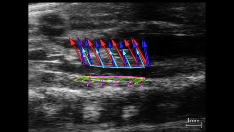
Abdominal Aorta Used In Strain Analysis
B-mode image of the abdominal aorta with vessel wall velocity vector arrows used in strain analysis.
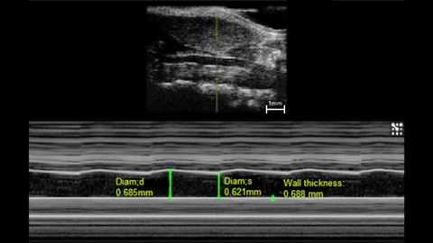
M-Mode of a Mouse Carotid Artery
M-mode image of mouse left common carotid.
Measurements: wall thickness and vessel diameter in systole and diastole.
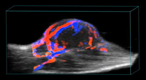
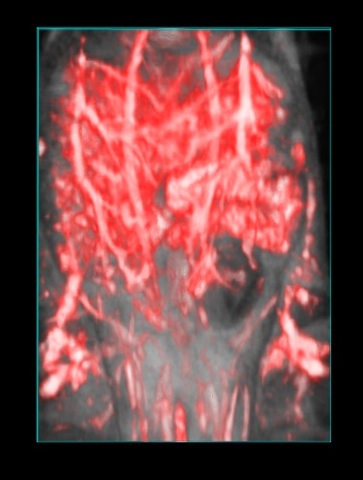
Splenic Vasculature in a Mouse
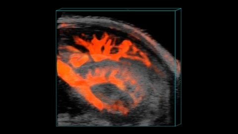
Murine Renal Vasculature in 3D
3D Spleen and Kidney Vasculature
3D Power Doppler rendering of a mouse spleen and kidney, highlighting splenic and renal vasculature.

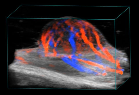
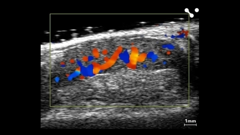
Blood flow in tumor
3D rendered high-resolution ultrasound (greyscale) and color Doppler (orange and blue) image of a subcutaneous tumor showing blood flow.
Blood flow in tumor
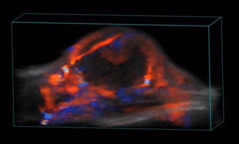
Blood flow in tumor
3D rendered high-resolution ultrasound (greyscale) and color Doppler (orange and blue) image of a subcutaneous tumor showing blood flow.
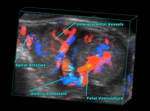
Mid-gestational Mouse Fetus and Placenta
3D color Doppler rendering of a mid-gestational mouse fetus and placenta, highlighted placental and fetal vasculature.
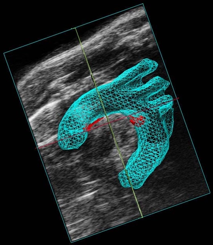
Mouse Aortic Arch
Color Doppler image of the aortic arch in a mouse showing all three branches.
Blood flow in tumor
3D rendered high-resolution ultrasound (greyscale) and color Doppler (orange and blue) image of a subcutaneous tumor showing blood flow.
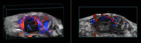
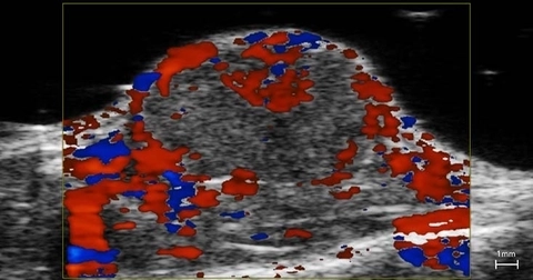
Blood flow in orthotopic tumor
3D rendered (left) and 2D (right) high-resolution ultrasound (greyscale) and color Doppler (orange and blue) image of an orthotopic pancreatic tumor showing blood flow.
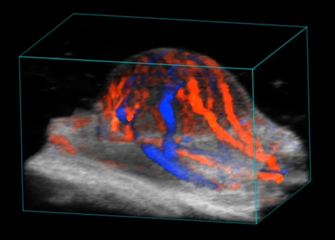
Blood flow in tumor
3D rendered high-resolution ultrasound (greyscale) and color Doppler (orange and blue) image of a subcutaneous tumor showing blood flow.
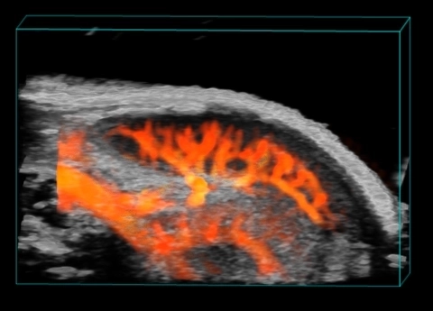
Vasculature and gut in the mouse abdomen
3D rendered photoacoustic (red) and ultrasound (greyscale) image of the mouse abdomen. Vasculature and gut contents can be seen in the photoacoustic image and are overlaid on the anatomical ultrasound image.
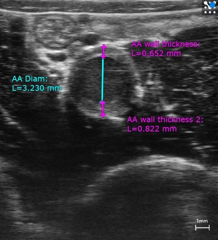
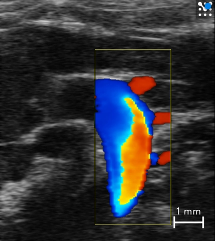
Rabbit Abdominal Aorta
Abdominal aorta in a rabbit imaged in B-Mode with thickened walls highlighted.
Atherosclerosis in the Aortic Arch
3D volume reconstruction of the mouse aortic arch (in blue) with atherosclerotic plaques (in red).