Liver
Make your Hepatic Research Robust with Vevo Ultra-High Frequency (UHF) Ultrasound Imaging
With the Vevo UHF imaging systems you can delineate organs, abnormalities, and tumors with high resolution.
Users can also assess functionality within the liver using the Color and PW Doppler (assess blood flow velocity) and 3D tools.
Watch Liver & Nash video On Demand.
How Can the Ultra-High Frequency Ultrasound Help You?
- Non-invasive, pre-palpable tumor identification
- Liver/tumor perfusion and volume quantification
- Contrast-enhanced imaging of biomarkers
- Assess and quantify oxygenation/hypoxia
Great for:
- Liver metastasis models
- Liver Cirrhosis (obtain channel/RF data for offline development)
- Non-alcoholic fatty liver disease
- Non-alcoholic steatohepatitis (NASH)
Do you have a question?
Recorded Webinar
Ultrasound of Liver Fibrosis & Hepatocellular Carcinoma
Presenter: Laith Sultan, Post Doctoral Fellow in the Department of Radiology Ultrasound Research Lab at the University of Pennsylvania.
Includes the following and more:
- Multiparametric quantitative B-Mode ultrasound for monitoring liver fibrosis
- Quantitative multimodality assessment of Antivascular Ultrasound (AVUS) therapy for hepatocellular carcinoma (HCC)
Recorded Webinar
Quantitative Ultrasound and Photoacoustic Imaging of Liver Fibrosis
Presented by Laith R. Sultan MD MPH and Mrigendra B. Karmacharya PhD from the PENN Ultrasound Lab at the University of Pennsylvania, on March 11th, 2021.
Includes the following and more:
- Liver fibrosis as public and global health issue
- The advantages and disadvantages of the current imaging and non-imaging diagnostic tools of liver fibrosis.
Image Gallery

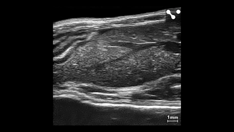
Shear Wave Elastography: Wide Box in Healthy Liver
Showing the wide SWE box used in a healthy mouse liver on the UHF29x.


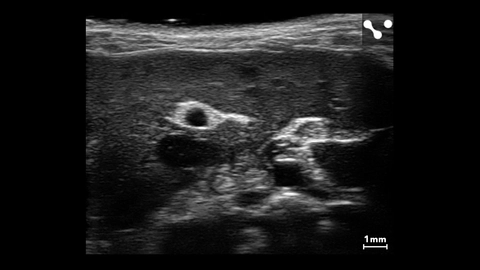
Right Liver Lobe and Adrenal Gland
B-mode image of the right liver lobe and adrenal gland, acquired using a UHF57x transducer on the Vevo F2.
Mouse Liver with Major Vessels
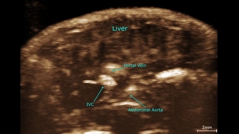
Vascular perfusion in the mouse liver
Mouse liver contrast MIP scanned using a UHF29x transducer.
Liver Vasculature in a Mouse
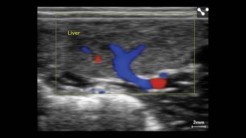
Liver Color Doppler in Small Dog
Liver Color Doppler of small dog scanned using a L38xp transducer.
Publications
TOP PAPER
Endothelial cells drive organ fibrosis in mice by inducing expression of the transcription factor SOX9
Science Translational Medicine
,
TOP PAPER
Generation of liver metastases in a mouse model using ultrasound-guided intravenous injection
STAR Protocols
,
TOP PAPER
Assessment of Transarterial Chemoembolization Using Super-resolution Ultrasound Imaging and a Rat Model of Hepatocellular Carcinoma
Ultrasound in Medicine & Biology
,
TOP PAPER
Quantitative Functional Evaluation of Liver Fibrosis in Mice with Dynamic Contrast-enhanced Photoacoustic Imaging
Radiology
,
TOP PAPER
Photoacoustic Imaging for Assessing Tissue Oxygenation Changes in Rat Hepatic Fibrosis
Diagnostics
,
TOP PAPER
Scattering Signatures of Normal versus Abnormal Livers with Support Vector Machine Classification
Ultrasound in Medicine & Biology
,
TOP PAPER
B-mode ultrasound for the assessment of hepatic fibrosis: a quantitative multiparametric analysis for a radiomics approach
Scientific Reports
,
TOP PAPER
Imaging-based vascular-related biomarkers for early detection of acetaminophen-induced liver injury
Theranostics
,
TOP PAPER
Noninvasive monitoring of liver metastasis development via combined multispectral photoacoustic imaging and fluorescence diffuse optical tomography
International Journal of Biological Sciences
, Request a Quote or Demo