Pulmonary
Discover the Benefits of Lung Ultrasound for Small Animals
Vevo ultra-high frequency ultrasound provides a fast, non-invasive method for identifying pulmonary pathologies.
Including:
- Pleural effusion
- Pleural thickening
- Pleural defects
- White lung
- Heart failure with preserved ejection fraction
Our technology is also an ideal option for non-invasive induction of your lung cancer models using image-guided injection.
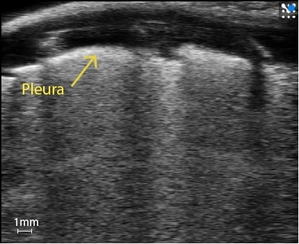
Healthy Lung Characteristic Artifacts:
- Bright pleural line at top
- A-lines - Curved, bright lines between dark rib shadows
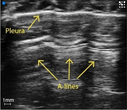
Image courtesy of: M.Sc. Niklas Hegemann, Kübler lab, Institute of Physiology, Charité-Universitätsmedizin Berlin & Dr. Jana Grune, Nahrendorf lab, Center for Systems Biology, Massachusetts General Hospital.
Do you have a question?
Diseased Lung

Pleural thickening
Pleural thickening can be observed.
Image courtesy of M.Sc. Niklas Hegemann, Kübler lab, Institute of Physiology, Charité-Universitätsmedizin Berlin & Dr. Jana Grune, Nahrendorf lab, Center for Systems Biology, Massachusetts General Hospital.

Lung infection
Lung infection can be detected and compared to healthy lung tissue.

Pleural effusion
Pleural effusion can be seen along with B-lines coming down from the pleura.
Image courtesy of M.Sc. Niklas Hegemann, Kübler lab, Institute of Physiology, Charité-Universitätsmedizin Berlin & Dr. Jana Grune, Nahrendorf lab, Center for Systems Biology, Massachusetts General Hospital.

White lung
In more severe cases numerous B-lines appear together as white lung (observed in interstitial-alveolar sydrome.
Image courtesy of M.Sc. Niklas Hegemann, Kübler lab, Institute of Physiology, Charité-Universitätsmedizin Berlin & Dr. Jana Grune, Nahrendorf lab, Center for Systems Biology, Massachusetts General Hospital.
Gallery
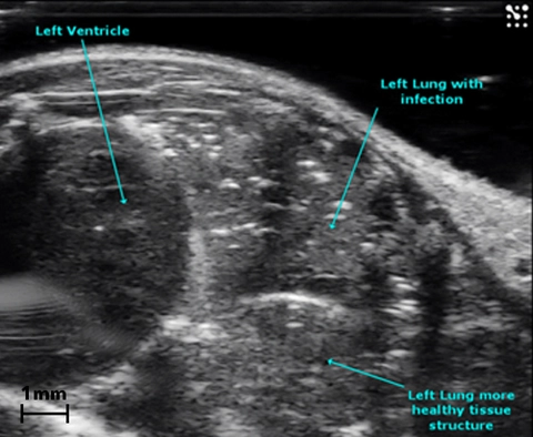
Lung Infection
B-Mode image of the mouse thorax illustrating healthy lung tissue and infected lung tissue.
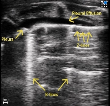
Rat Pleural Effusion
Rat with pleural effusion and pleural thickening. Clear B-Lines can be seen as well as Z-Lines.
Image courtesy of : M.Sc. Niklas Hegemann, Kübler lab, Institute of Physiology, Charité-Universitätsmedizin Berlin & Dr. Jana Grune, Nahrendorf lab, Center for Systems Biology, Massachusetts General Hospital.
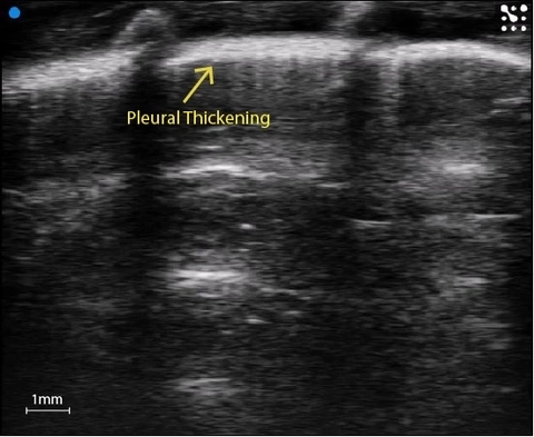
Pleural thickening
Substantial pleural thickening in the rat lung.
Image courtesy of : M.Sc. Niklas Hegemann, Kübler lab, Institute of Physiology, Charité-Universitätsmedizin Berlin & Dr. Jana Grune, Nahrendorf lab, Center for Systems Biology, Massachusetts General Hospital.
Murine White Lung
Mouse lung covered in B-Lines, creating white lung.
Image courtesy of : M.Sc. Niklas Hegemann, Kübler lab, Institute of Physiology, Charité-Universitätsmedizin Berlin & Dr. Jana Grune, Nahrendorf lab, Center for Systems Biology, Massachusetts General Hospital.

Healthy mouse lung
Healthy naive mouse lung with echogenic pleura and A-Lines ringing down.
Image courtesy of : M.Sc. Niklas Hegemann, Kübler lab, Institute of Physiology, Charité-Universitätsmedizin Berlin & Dr. Jana Grune, Nahrendorf lab, Center for Systems Biology, Massachusetts General Hospital.
Publications
TOP PAPER
Endothelial cells drive organ fibrosis in mice by inducing expression of the transcription factor SOX9
Science Translational Medicine
,
TOP PAPER
Lung ultrasound as a translational approach for non-invasive assessment of heart failure with reduced or preserved ejection fraction in mice
Cardiovascular Research
,
TOP PAPER
High Resolution Ultrasound and Photoacoustic Imaging of Orthotopic Lung Cancer in Mice: New Perspectives for Onco-Pharmacology
PLOS ONE
, Comprehensive Ultrasound Imaging of Right Ventricular Remodeling Under Surgically Induced Pressure Overload in Mice
American Journal of Physiology-Heart and Circulatory Physiology
, Pyruvate kinase M2 activation protects against the proliferation and migration of pulmonary artery smooth muscle cells
Cell and Tissue Research
, Lung ultrasound is a reliable method for evaluating extravascular lung water volume in rodents
BMC Anesthesiol
, Transplacental sildenafil rescues lung abnormalities in the rabbit model of diaphragmatic hernia
Thorax
, Request a Quote or Demo