Pancreas
A High-resolution, Multi-modal Solution for Robust Evaluation of Small Animal Pancreatic Tumor Models
From model generation with image-guided injection to characterization of the tumor micro-environment.
Vevo ultra-high frequency ultrasound and photoacoustic technology enables you to get a complete understanding of the anatomy and physiology in your specific tumor models and monitor response to treatment longitudinally.
With Vevo Imaging Systems you can:
- Non-invasively inject and generate orthotopic models with precision
- Quickly and accurately measure tumor size
- Monitor perfusion kinetics with contrast enhanced ultrasound
- Characterize tumor micro and macro vasculature in response to treatment
- Assess intratumoral hypoxia
- Perform targeted measurements of nanomedicines or theranostic agents
Do you have a question?
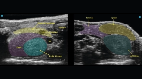
Healthy Pancreas
Anatomical location of the healthy mouse pancreas with ultrasound. The head of the pancreas is seen on the left and the tail of the pancreas can be visualized on the right.
Image-guided Needle Injection
Example of image guided injection into the tail of the mouse pancreas. This technique can be used to deliver cancer cells to develop a model, as a non-invasive alternative to surgery.
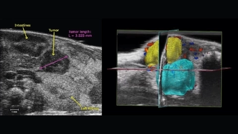
Pancreatic Tumor
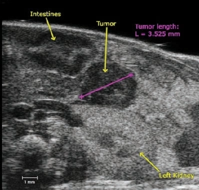
Left: Small pancreatic tumor in a mouse model with approximate sizing in purple.
Right: 3D Color Doppler image of a pancreatic tumor (yellow) and the kidney (blue) with volumetrics performed.
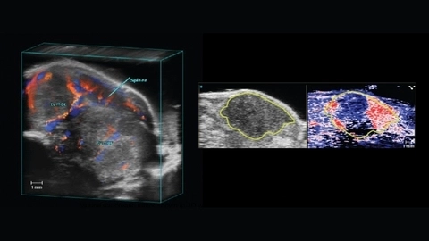
3D and B-Mode
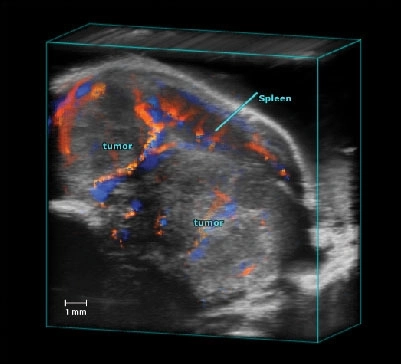
Left: Murine pancreatic tumor shown in 3D with color Doppler highlighting major vasculature going away from the transducer (blue) and towards the transducer (red).
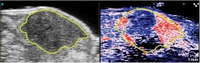
Right: B-Mode image of a murine pancreatic tumor (circled in yellow) on left with co-registered photoacoustic Oxy-Hemo Mode on right. Red depicts oxygenated hemoglobin and blue depicts deoxygenated hemoglobin.
Gallery

Left Kidney, Spleen and Pancreas
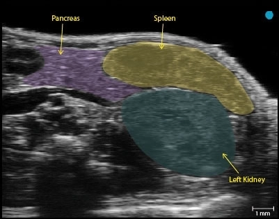
B-mode image of the left kidney, spleen and pancreas acquired using a UHF57x transducer on the Vevo F2.
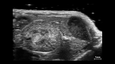
Mouse Kidney, Spleen and Pancreas

Pancreatic tumor oxygen saturation
Pancreatic tumor size
Pancreatic Tumor Color Doppler 3D
Healthy Pancreas - Tail
Publications
TOP PAPER
Quantification of Murine Pancreatic Tumors by High-Resolution Ultrasound
Methods in Molecular Biology
, Ketogenic diet and chemotherapy combine to disrupt pancreatic cancer metabolism and growth
Med
, T Cell–Mediated Antitumor Immunity Cooperatively Induced by TGFbR1 Antagonism and Gemcitabine Counteracts Reformation of the Stromal Barrier in Pancreatic Cancer
Molecular Cancer Therapeutics
, PEGPH20, a PEGylated human hyaluronidase, induces radiosensitization by reoxygenation in pancreatic cancer xenografts. A molecular imaging study
Neoplasia (United States)
, Method To Visualize the Intratumor Distribution and Impact of Gemcitabine in Pancreatic Ductal Adenocarcinoma by Multimodal Imaging
Analytical Chemistry
, Interrogating the immune-modulating roles of radiation therapy for a rational combination with immune-checkpoint inhibitors in treating pancreatic cancer
Journal for ImmunoTherapy of Cancer
, Selective Alanine Transporter Utilization Creates a Targetable Metabolic Niche in Pancreatic Cancer
Cancer discovery
, Immunoevolution of mouse pancreatic organoid isografts from preinvasive to metastatic disease
Scientific Reports
, Request a Quote or Demo