Color Doppler
Visualize Flow Velocities Within Vessels to Monitor Direction of Flow

Easily Identify Blood Vessel Structures

Color doppler mode provides your “road map” – a perfect guidance system

Color Doppler Image Illustrating Vasculature in the Mid-gestation Mouse Placenta

Using Color Doppler Mode, flow velocities within vessels are color-coded!

Vessel networks and flow patterns can be laid out in a complex manner. Using Color Doppler, the intensity of the color is a function of velocity. Flow towards and away from the transducer is indicated either as red or blue, respectively. To remember this, use the name of a famous yellow cartoon character as an acronym: BART – Blue Away, Red Towards (with the top of the screen as your reference point).

Benefits of the Color Doppler Mode:

  • Provides a visual overview of flow within the vessel or heart
  • Rapid identification of vessels, valves, turbulent flow
  • Assess flow direction and velocity
  • Quantify volume and percent vascularity when combined with 3D Mode
  • Guidance for reproducible quantification of flow velocities using Pulsed-Wave Doppler

When should you use the Color Doppler Mode?

  • Identify area of stenosis or thrombosis (e.g. TAC or aneurism models)
  • Determine the presence and amount of arterial plaques and associated turbulent flow
  • Find small vessels such as mouse coronary arteries, femoral and arcuate arteries
  • Evaluate blood flow after a stroke or other conditions causing impaired blood flow
  • Monitor blood flow to major organs such as heart, kidney, liver pancreas, carotid, abdominal aorta, and others

Examples of Color Doppler

Tumor-Colour-Doppler
Subcutaneous tumor

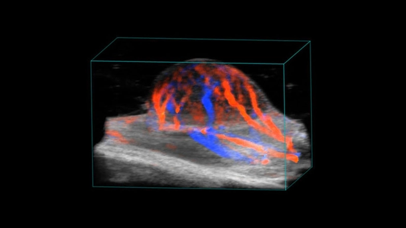
3D color doppler image of a subcutaneous tumor.

Color Placenta and Embryo
Mid-gestation Mouse Placenta

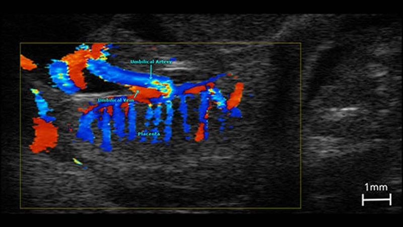
Color doppler image illustrating vasculature in the mid-gestation mouse placenta.

Want to learn more?