MSK
See changes in tissue and vascular structure in 2D & 3D
How much inflammation is in the arthritic joints? Assess the musculoskeletal system with Vevo technology!
Using ultra-high frequency ultrasound and photoacoustics, visualize changes in tissue and vascular structure in 2D and 3D with resolution down to 30 microns, in vivo. Tissue oxygenation, capillary function, tissue inflammation, bone erosion and more can be assessed longitudinally without interfering with your experiment.
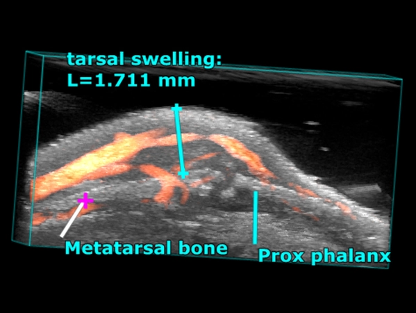
3D power Doppler image of a swollen joint due to collagen-induced arthritis. Image courtesy of Dr. Mandl, University of Vienna, Austria.
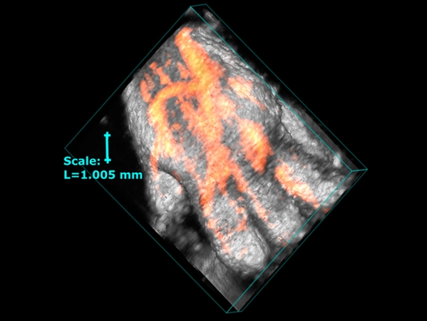
3D image illustrating the vascularity in the mouse paw in a model of collagen-induced arthritis. Image courtesy of Dr. Mandl, University of Vienna, Austria.
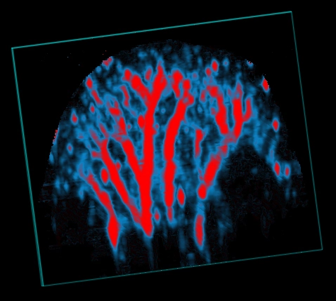
Multiplexed photoacoustic image of the mouse hindlimb after ICG injection (isolated in the popliteal lymph node).
3D rendered image of oxygen saturation in the mouse ear, imaged with photoacoustics in Oxy-Hemo Mode.
• Collagen induced arthritis (CIA)
• TNFa overexpressing animals
• Musculoskeletal (MSK) disease
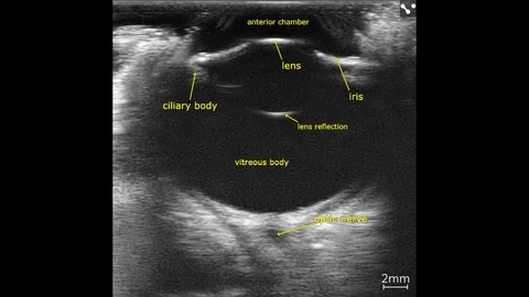
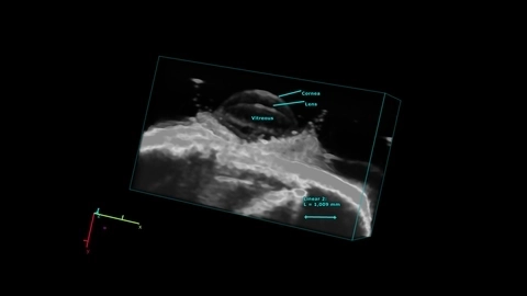
Entire canine eye visualized in vivo with the Vevo F2, scanned using a UHF22x transducer.
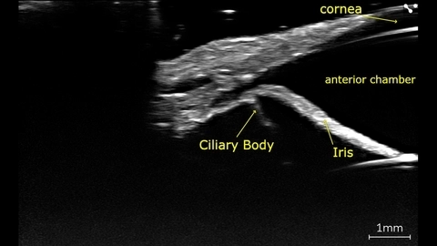
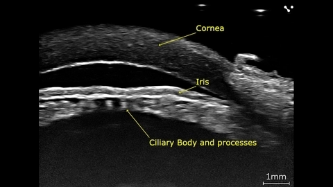
Canine eye imaged in vivo on the Vevo F2 scanned using the UHF71x.
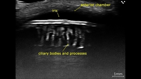
In vivo canine eye with ciliary processes, imaged with high-frequency on the Vevo F2 scanned using the UHF71x.
Porcine eye imaged ex-vivo using the Vevo F2 with a UHF71x transducer.
Mouse eye scanned using the UHF71x transducer using the Vevo F2 in 3D.
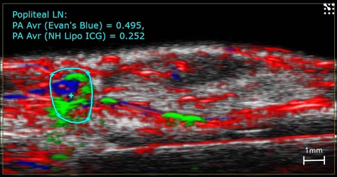
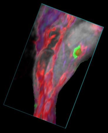
High-resolution ultrasound (greyscale) and photoacoustic (red, blue and green) image of the mouse hindlimb showing Evan's Blue (blue) and ICG (green) dye in the lymphatic vessels and the popliteal lymph node.