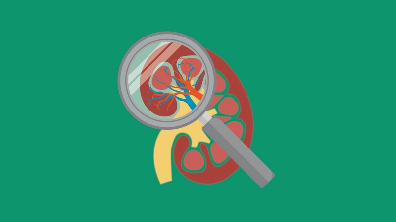
Ultra High-Frequency Ultrasound
Pioneering the Use of Ultra-High Frequency Ultrasound in Medical Research

Conventional vs. Ultra-high Frequency Ultrasound
Ultra-high Frequency Ultrasound Technology Available for Preclinical Research
The Vevo imaging platform was the world’s first commercially available ultra-high frequency array-based ultrasound imaging system and has since emerged as the gold standard in small animal anatomical and functional in vivo imaging.
The Vevo ultra-high frequency ultrasound technology enables the researcher to obtain in vivo anatomical, functional, physiological and molecular data simultaneously, in real-time and with a resolution down to 30 µm. The systems are easy to use, non-invasive and fast—providing extremely high throughput when needed.
They are designed with the researcher in mind, with system presets and animal handling tools for fast image acquisition and numerous protocols, software and data management tools optimized for today’s scientists. Learn more about our ultra high frequency ultrasound systems for preclinical research.
How does it work?
Tissues of different densities absorb and reflect sound waves differently. Reflection of the sound waves occurs at the boundaries between tissues of differing densities (differences in acoustic impedance). The reflected (echoed) ultrasound waves are detected by the “listening” transducers and the information is converted into electrical signals that are used to create an ultrasound image.
The magnitude of the reflected waves will be greater as the difference in acoustic impedance increases, which will result in a brighter (more echogenic) image. Detecting these differences is what forms the differences in grey-scale in a typical B-Mode ultrasound image. The resolution achieved on ultrasound systems depends on the frequency of the ultrasound waves being emitted.
Our ultra high frequency ultrasound systems give you high resolution—down to 30 µm while imaging depths up to 3cm at frame rates up to 10,000 fps.
Ultra-High Frequency, Now at the Point of Care
Clinicians can now harness the power of 46 MHz ultra-high frequency ultrasound beyond the research lab. Our parent company, FUJIFILM Sonosite, now offers the first and only 46 MHz transducer built specifically for point-of-care, available exclusively on Sonosite LX. Designed to provide detailed imaging of submillimeter anatomy in the first centimeter of the imaging field, this novel solution delivers the best superficial imaging available in POCUS today.
With UHF46-20, Sonosite LX has the largest frequency range on any POCUS system, combining standard frequencies with ultra-high frequency in one device.
Get information on:
- Blood flow, localization, and direction
- Tissue motion over time
- Presence of molecular biomarkers
- Anatomy and size of 2D, 3D and 4D regions
- Cardiac and vascular strain
Key Benefits of High-Frequency Ultrasound for your in vivo Research
High Resolution
Visualize anatomical function in high resolution; detail like you've never seen before using ultrasound. Co-registration of anatomical, functional, physiological and molecular data.

In vivo, In Real Time
Study high speed events such as blood flow and cardiac function as it happens in living organisms. Real-time detection of anomalies. In vivo imaging for better translation to the clinic.

Safe, Non-invasive and Non-radioactive
No negative side effects. UHF ultrasound supports the 3Rs of animal use: replacement, reduction and refinement.

Longitudinal and Cost-efficient
Monitor the disease in the same subject over time, in vivo. UHF ultrasound is a more cost-effective imaging solution when compared with other modalities like MRI/PET.