Liver Gallery

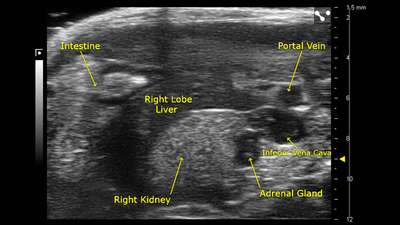
Right Liver Lobe and Adrenal Gland
B-mode image of the right liver lobe and adrenal gland, acquired using a UHF57x transducer on the Vevo F2.
Mouse Liver with Major Vessels
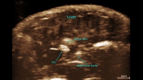
Vascular perfusion in the mouse liver
Mouse liver contrast MIP scanned using a UHF29x transducer.
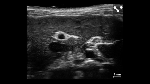
Liver Vasculature in a Mouse
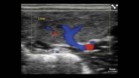
Liver Color Doppler in Small Dog
Liver Color Doppler of small dog scanned using a L38xp transducer.
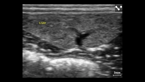
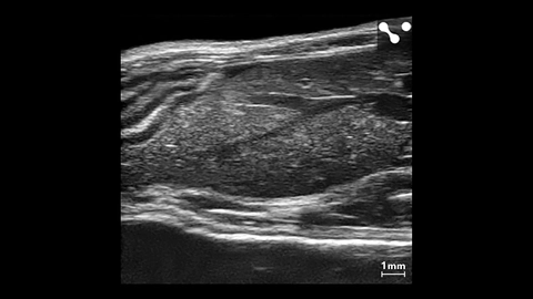
Liver of Small Dog
Liver of small dog measured using a L38xp transducer.

Liver Metastasis
Cross section of the mouse abdomen with liver metastasis.
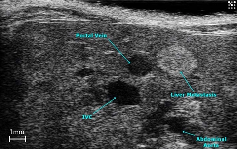
Liver Metastasis
Cross section of the mouse abdomen highlighting major structures, including liver metastasis.
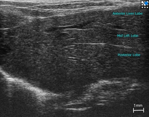
Lobes in the Mouse Liver
Mouse liver with lobes labelled.
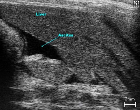
Ascites in the Rat Liver
B-Mode image of ascites in a cirrhotic rat liver.
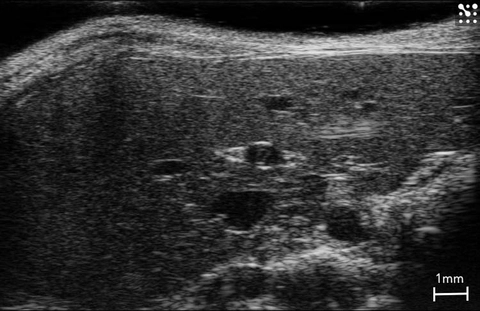
Mouse Liver
Healthy mouse liver imaged in B-Mode.