Vascular
Visualize the Smallest Vascular Anatomy Possible
The Vevo MD was designed specifically with vascular applications in mind.
Ultra high frequency vascular ultrasound allows for visualization of the smallest vascular anatomy imaginable:
- Arteries and veins in pediatric and neonatal patients
- Measurement of Intima-Media Thickness (IMT) for research and assessment of cardiovascular health
- Assessment of vein wall morphology for cannulation readiness in AV fistula patients
- Assessment of peripheral vessels in diabetes and other circulatory conditions
- Visualization of flow patterns in in atherosclerotic or abnormal vessels
Gallery
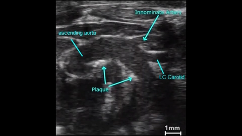
Atherosclerotic Plaque in an Aortic Arch
2D image showing atherosclerotic plaques in a mouse aortic arch.

Radial Artery - Adult Female

Publications
TOP PAPER
Ultra-High Frequency Ultrasound, A Promising Diagnostic Technique: Review of the Literature and Single-Center Experience
Canadian Association of Radiologists Journal
, Validity of ultrasound-guided identification of dilated lymphatic vessels to detect early breast cancer-related lymphedema
Archives of Plastic Surgery
, Tomographic ultrasound for three-dimensional visualization of temporal arteries
Scandinavian Journal of Rheumatology
, Quantitative ultrasound and photoacoustic assessments of red blood cell aggregation in the human radial artery
Photoacoustics
, Investigating the presence and detectability of structural peripheral arterial changes in children with well-regulated type 1 diabetes versus healthy controls using ultra-high frequency ultrasound: a single-centre cross-sectional and case-control study
eClinicalMedicine
, Impact of kidney function on stiffness of small conduit arteries in hypertension and obesity
Journal of Hypertension
, Vascular structure and stiffness in pediatric Mulibrey nanism using ultra-high frequency ultrasound
Veins and Lymphatics
, Tube-in-Tube Phalloplasty with Tailor-made Bilateral Superficial Circumflex Iliac Artery Perforator Flaps Using Preoperative High-resolution Ultrasound
Plastic and Reconstructive Surgery - Global Open
, Deep Fat Saving Elevation of the Superficial Circumflex Iliac Artery Perforator Flap
Medicina (Lithuania)
, Request a Quote or Demo