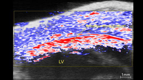
Anterior Myocardium Oxygenation, High O2 stats in Red, Low in Blue
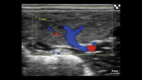
Liver Color Doppler in Small Dog
Liver Color Doppler of small dog scanned using a L38xp transducer.
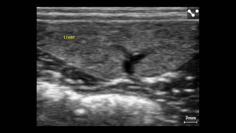
Liver of Small Dog
Liver of small dog measured using a L38xp transducer.
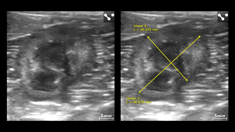
Left Kidney in Small Dog with Measurements
Left kidney in small dog with measurements imaged using the L38xp transducer.
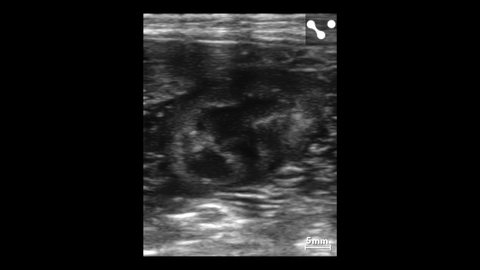
Kidney in a Small Canine
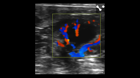
Kidney Color Doppler in Small Dog
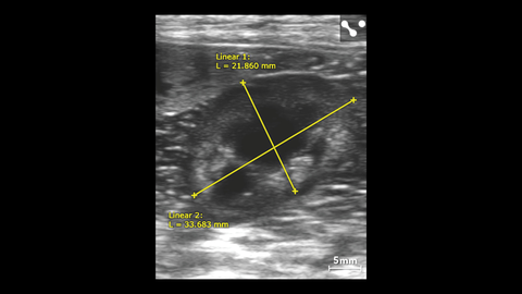
Canine Left Kidney with Measurements
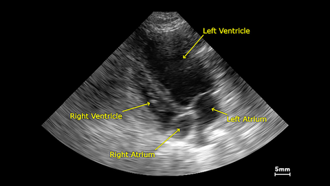
Apical View in a Small Dog
Aortic Arch in a Mouse
Ultrasound guided free hand injection into the murine pancreas
Acquired using the Vevo 3100.
40uL volume injected into the mouse pancreas transcutaneously with image-guided injection
Acquired using the Vevo 3100.
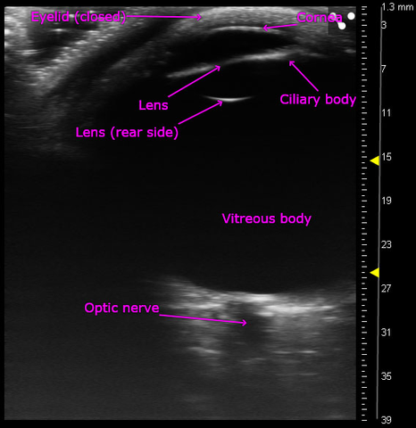
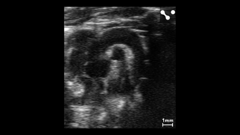
Entire adult human eye images with ultra high frequency ultrasound
Acquired using the Vevo MD.
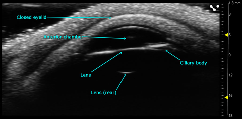
Anterior chamber of the adult human eye
Acquired using the Vevo MD.
Brain vasculature visualized using Contrast Microbubbles
Infusion of contrast microbubbles into the brain microvasculature of a mouse.
Vasculature in the Brain during Ischemic Stroke
Vasculature in the mouse brain, visualized with color Doppler during transient carotid artery occlusion.
Vasculature in the Mouse Brain
Microvasculature in the mouse brain visualized in 4D after contrast microbubble injection.
Mid-gestational Mouse Placenta
Power Doppler imaging showing vascularization in the various regions of the placenta.
Placental Oxygen Saturation
Co-registered high-resolution ultrasound (greyscale) and photoacoustic (red, white and blue) image of a mid-gestation embryo (left) and its associated placenta. The photoacoustic signal depicts oxygen saturation, with red representing regions of higher sO2.
Kidney Vessel Branching
3D Power Doppler showing branching of the splenic artery and kidney vessels in a mouse.
Mouse Kidney Vasculature
Power Doppler image of mouse kidney vasculature acquired using the UHF46x probe.
Mouse Brain Vasculature
3D Power Doppler rendering of a mouse brain, highlighting cerebral vessels.