Congratulations!
Thank you to all those that entered our Seeing More Matters image contest. We received a lot of great submissions from around the world. Entries were judged on the following:
- Subject matter (application/animal model)
- Quality of image (clarity)
- Details that are visible (visualization of subject matter)
- Innovative use of the imaging system
We are happy to announce the winners below:
1st Place Winner:
Kelsey Kubelick, Georgia Institute of Technology, USA. System used: Vevo LAZR
Kelsey will receive the iPad Mini for her entry below.

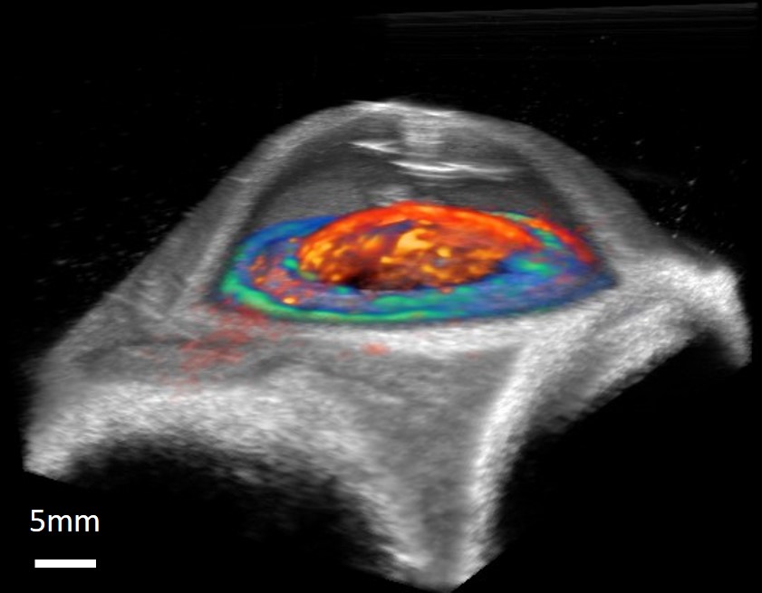
Description: Combined ultrasound/photoacoustic image of a porcine eye after delivering stem cells labeled with gold nanospheres. Ultrasound shows the needle track at the top of the cornea after injecting stem cells. Spectroscopic analysis separates photoacoustic signals from endogenous absorbers and labeled stem cells. The photoacoustic signal from gold nanosphere-labeled stem cells is colored orange. Stem cells cover the lens and iris at 5 hours post-injection. The photoacoustic signal from the iris, due to melanin absorbance, is colored blue. Kelsey also submitted a video: See video submission here.
Second Place Winner:
Jonathan Lavaud
Institute for Advanced Biosciences/OPTIMAL platform, ,France
System used: Vevo LAZR
Wins a VisualSonics gift bag of branded items.
Third Place Winner:
Jeroen Molinger
Erasmus MC
Netherlands
System used: Vevo MD
Wins a VisualSonics bag of gift branded items.
Title: Multimodal Melanoma brain metastasis
[[{"fid":"3858","view_mode":"default","fields":{},"type":"media","field_deltas":{"3":{}},"attributes":{"class":"media-element file-default","data-delta":"3"}}]]Title: Radial Artery Thrombosis
[[{"fid":"3860","view_mode":"default","fields":{},"type":"media","field_deltas":{"4":{}},"attributes":{"class":"media-element file-default","data-delta":"4"}}]]Description: Orthotopically implanted melanoma brain metastasis imaged with VEVO-Lazr photoacoustic imaging system (Tumoral hemoblonin content; OXY-hemo protocol) merged with CT anatomical image. The fusion of both modalities was based on the anatomical structure of pure ultrasound informations gived by the VEVO-Lazr system. Click to enlarge.
Description: Radial artery thrombosis after line insertion. Link to video
To view all entries, click here.