Vevo F2 Gallery
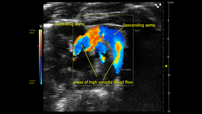
Color Doppler of Mouse Aortic Arch
Color Doppler image of an aortic arch in a female mouse, acquired using a UHF57x transducer on the Vevo F2.
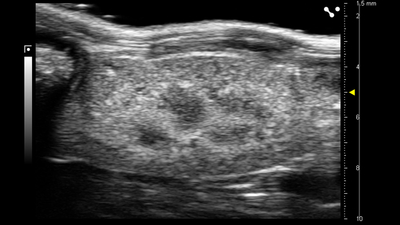
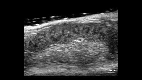
Sagittal Kidney
B-mode image of the sagittal kidney acquired using a UHF57x transducer on the Vevo F2.
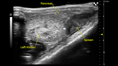
Left Kidney, Spleen and Pancreas
B-mode image of the left kidney, spleen and pancreas acquired using a UHF57x transducer on the Vevo F2.

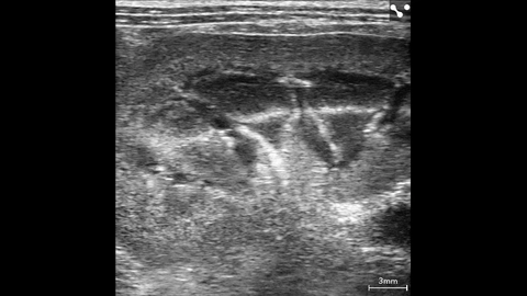
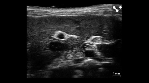
Lymph nodes in rhesus macaque
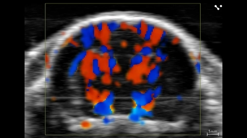
Juvenile rat cerebral vasculature
Splenic Vasculature in a Mouse
Color Doppler of Rat Kidney Vasculature
Color Doppler of rat kidney vasculature scanned using a UHF29x transducer.
Rabbit Kidney
Rabbit kidney scanned using a UHF29x transducer.
Murine Spleen
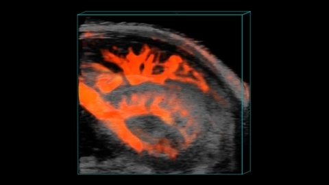
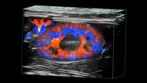
Murine Renal Vasculature in 3D
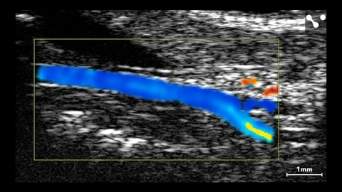
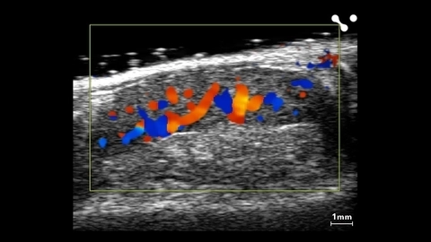
Murine Carotid Artery
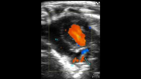
Murine Apical Color Doppler
Murine apical color Doppler scanned using a UHF46x transducer.
Mouse Liver with Major Vessels