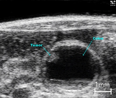
Colon Cancer

B-Mode image of the mouse colon in cross-section with presence of a tumor highlighted.
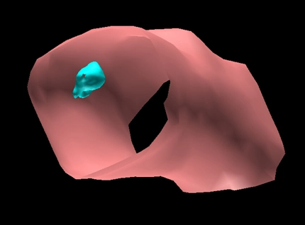
3D Surface Volume of the Mouse Colon with Tumor

Surface view of a 3D volume of a section of mouse colon (in pink) with a tumor (in blue).

Liver Metastasis

Cross section of the mouse abdomen with liver metastasis.

Tumor microvascularization
Intravenous bolus injection of non-targeted MicroMarkerTM contrast agent in a mouse mammary tumor. The MIP(Maximum Intensity Persistence) setting can be adjusted to give a better visual representation of the vascular tree.

Liver perfusion MIP Processed
Advanced contrast quantification of liver perfusion in color coded parametric map with the VevoCQTM software analysis tool.

Liver perfusion
Contrast-enhanced ultrasound show any selective enhancement in the venous and arterial phases.

Kidney Microvascularization
Intravenous bolus injection of non-targeted MicroMarkerTM contrast agent in a mouse left kidney. The MIP(Maximum Intensity Persistence) setting can be adjusted to give a better visual representation of the vascular tree.

GNR Post Injection
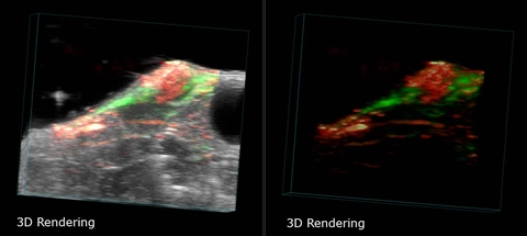
Breast Cancer Tumor in 3D

Breast Cancer Tumor in a mouse, imaged with photoacoustics in 3D. Shows localization of a targeted nanoparticle (green) within the tumor. Left panel shows both photoacoustics and ultrasound images, overlaid. Right panel shows only the photoacoustics signal.
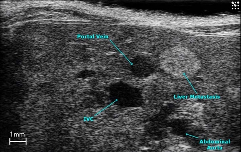
Liver Metastasis

Cross section of the mouse abdomen highlighting major structures, including liver metastasis.
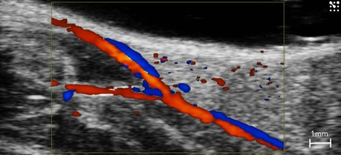
Femoral Artery Bifurcation

Bifurcation of the femoral artery in the mouse seen using color Doppler.

Vasculature in the Mouse Paw
Color Doppler image of the vasculature in the paw of a mouse.

Oxygen Saturation in the Mouse Paw
Oxy-hemo mode illustrating oxygen saturation in the mouse paw.

Knee Vasculature
Color Doppler image of vasculature of the mouse knee.
Image courtesy of Dr. Mandl, University of Vienna, Austria.

Metatarsal Phalangeal Joint in Arthritis
The Mouse Metatarsal Phalangeal Joint in a model of collagen-induced arthritis.

The Knee in Collagen-induced Arthritis
Anatomy of the mouse knee joint in a model of collagen-induced arthritis.
Image courtesy of Dr. Mandl, University of Vienna, Austria.
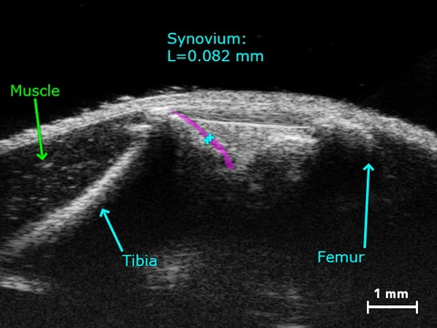
Synovium in a Healthy Knee

Synovium in a healthy mouse knee.
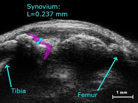
Synovium in Collagen-induced Arthritic Knee

Thickened synovium in the mouse knee due to collagen-induced arthritis.

Healthy Mouse Paw

Rendered 3D image of a healthy mouse paw.
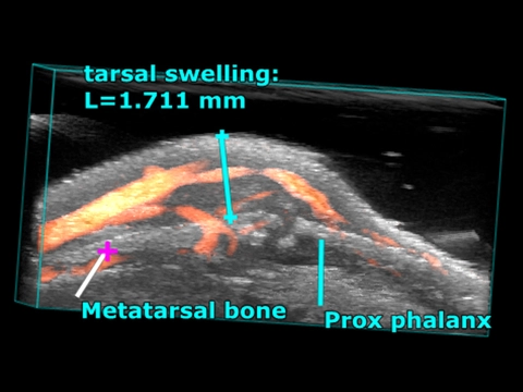
Arthritic Joint

Rendering of a 3D power Doppler image of a swollen joint due to collagen-induced arthritis.
Image courtesy of Dr. Mandl, University of Vienna, Austria.
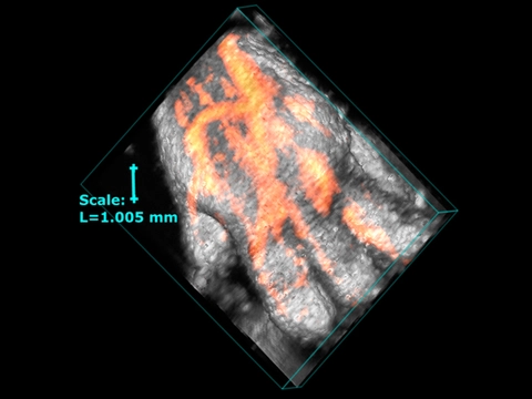
Vascularity in the Mouse Paw

Rendering of a 3D power Doppler image illustrating the vascularity in the mouse paw in a model of collagen-induced arthritis.
Image courtesy of Dr. Mandl, University of Vienna, Austria.