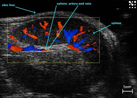
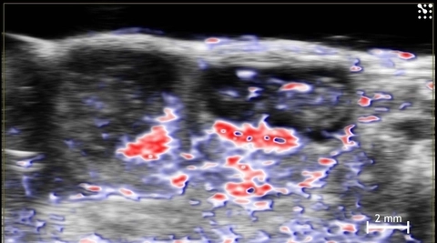
Splenic Vasculature
Color Doppler image of the mouse spleen and splenic vasculature.
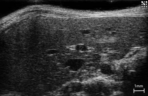
Mouse Liver
Healthy mouse liver imaged in B-Mode.

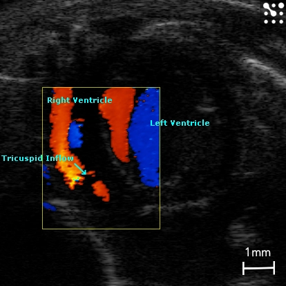
Tricuspid Flow in the Mouse
Apical four chamber view illustrating blood flow through the tricuspid valve, imaged with color Doppler.
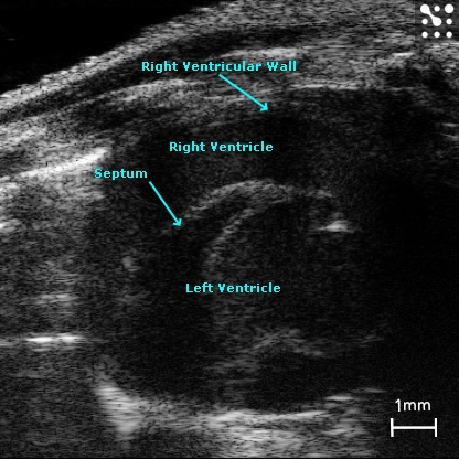
Mouse Right Ventricle
The right ventricle in a mouse imaged in B-Mode.
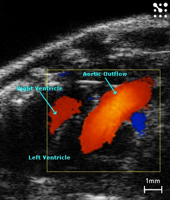
Ascending Aorta from a Parasternal Notch View
Color Doppler image showing blood flow through the ascending aorta and right ventricle from a parasternal notch view in a mouse.
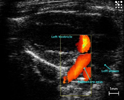
Left Atrium and Pulmonary Veins
Pulmonary veins entering the left atrium in the mouse; imaged with power Doppler.
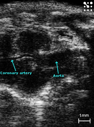
Mouse Coronary Artery
B-Mode image showing the mouse coronary artery branching of the root of the aorta.
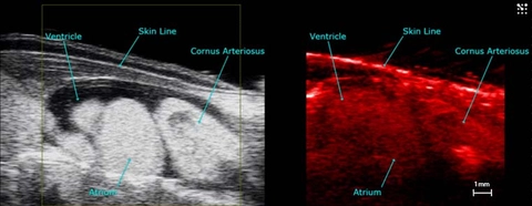
Axolotl heart in PA
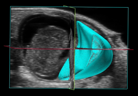
3D Reconstruction of the Placenta
3D reconstruction of the mid-gestational mouse placenta (in blue) with fetus to the left of the image.
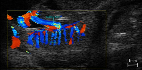
Placental Blood Flow
Color Doppler image illustrating blood flow in the fetus, umbilical cord and placenta in a late gestational mouse implantation site.
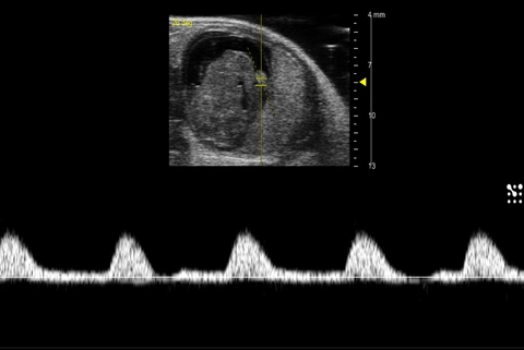
Uterine Artery Blood Flow
PW Doppler of the uterine artery in a mid-gestational mouse.
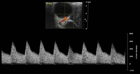
Umbilical Artery Blood Flow
PW Doppler of the umbilical artery in a mid-gestational mouse.
Oxygen Saturation in the Mouse Placenta and Fetus
Oxy-hemo phototacoustics image showing oxygen saturation in two mid-gestational mouse implantation sites. Red indicates oxygenated hemoglobin; blue indicates de-oxygenated hemoglobin.

Spiral Arteries in the Mouse Placenta
Color Doppler indicating direction on blood flow in placental spiral arteries in a mid-gestational mouse implantation site.