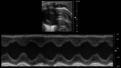
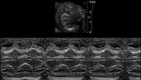
Short Axis M-Mode

Short axis M-Mode of a rat heart.

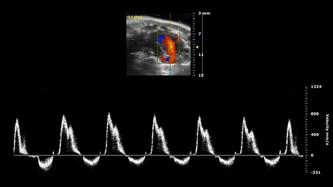
PW Doppler of Mitral Flow

PW Doppler of flow through the mitral valve from an apical 4 chamber view of the mouse heart with separation of E and A peaks visible.

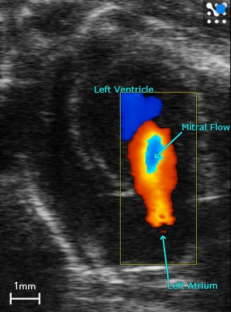
Apical 4 Chamber view

Apical 4 chamber view of the mouse heart with color Doppler indicating flow through the mitral valve.

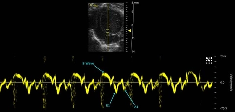
Mitral Tissue Doppler

Tissue Doppler of the mitral annulus in a mouse, showing common measurements to analyze diastolic function.

M-Mode of the Left and Right Ventricles in the Developing Mouse Heart


UHF 48 MHz - Radial Artery

UHJ 48 MHz - Arm Vein with Valve

UHF 70 MHz - Radial Artery

9UHF 48 MHz - Arm Vein Valve

Vevo MD UHF 70 - Superficial vein shows constriction of blood flow

Vevo MD UHF 48 Adult Radial Artery with Colour Flow

Vevo MD UHF 70 Superficial vein with low flow in adult

Vevo MD UHF 70 Adult Vein with Valves

Vevo MD UHF 70 Adult low flow vein 2D - See each blood cell passing through vessel

Vevo MD UHF 70 Superficial Vein with colour flow

Vevo MD UHF 70 Trx Artery and Vein 2D

Vevo MD UHF 70 Vein with Valves

Vevo MD UHF 48 - Radial artery in 1 month infant

Vevo MD UHF 48 Post tooth extraction palpable mass on a 40 Yr old male

Vevo MD UHF 48 Phantom with needle insertion
