The ultrasound of the bladder is a representative transabdominal view and clearly shows an infiltrating mass, albeit a small one, into the lumen of the bladder. This imaging plane was acquired using an MS550 transducer.
The layers of the esophageal wall can easily be distinguished using Vevo MD ultra high-frequency ultrasound. Physiology of the esophagus can also be observed while a volunteer swallows water.
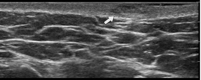
Vevo MD Ultra High-Frequency ultrasound is able to bring out details in scars that are not visible using conventional ultrasound. Here is an image of a scar formed after open heart surgery.
Here are a couple of awesome new applications for our brand new 4D Imaging Mode. Check out these videos showing incredible detail of the left atrium and right atrium!
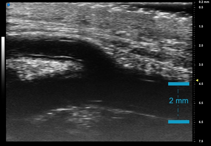
Although this may look like a carotid artery at first, what we are actually looking at is a longitudinal view of the radial artery bifurcation taken with our Vevo MD.
Our new 4D mode on the Vevo 3100 Imaging System will allow you to look at your 3D images over time. Here, a 4D image was taken from a parasternal short axis (SAX) view of the heart.