Oncology
Powerful, High Resolution Multi-modal Imaging Solution for Translational Cancer Research
Characterize tumors in vivo and non-invasively with ultra-high frequency (UHF) ultrasound
Conduct high-throughput screenings for tumor growth and detect and monitor growth and response of tumors to therapy at multiple time points in preclinical disease models all in vivo. In addtion, you can image the tumor microenvironment for changes and you have a powerful multi-modal platform for translational oncology research.
With the Vevo Imaging Systems you can:
- Non-invasively characterize tumor tissue in vivo
- Screen for very small lesions (resolution down to 30µm); early detection
- Accurately quantify volume of orthotopic and subcutaneous tumors longitudinally
- Perform rapid tumor volume assessments with a dedicated Oncology Measurement Package
- Visualize and quantify angiogenesis, vasculature and perfusion
- Measure hypoxia
- Assess biomarker or drug distribution
- Develop models using ultrasound-guided injections
Explore a Wide Range of Applications
A Comprehensive Solution for Pancreatic Cancer Researchers
VisualSonics offers you a complete imaging solution for orthotopic pancreatic cancer models, from model generation with image-guided injection to characterization of the tumor micro-environment.
Advance Cancer Research Using Molecular Imaging
Explore high-frequency ultrasound and photoacoustics for in vivo molecular imaging of targeted agents, theranostics and nanomedicine.
Vevo 4 Oncology Web Series
Pancreatic and Liver Tumor Imaging
Watch the first episode of the Vevo 4 Oncology Web Series, presented by Prof. Annarosa Arcangeli, and learn more about a powerful, high resolution multi-modal imaging solution for oncology. Now available on-demand!
Gallery
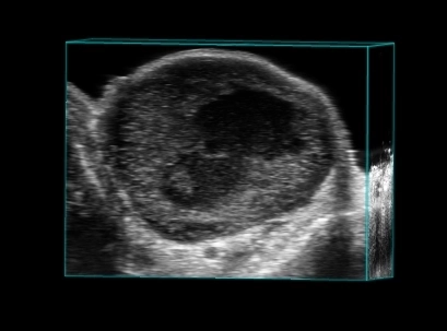
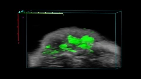
Breast tumor in 3D
Tumor 3D
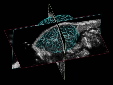
3D Volume Reconstruction of a Murine Tumor
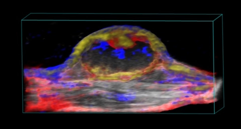
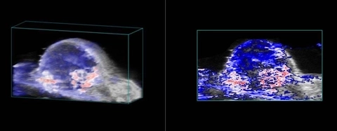
Nanoparticle distribution in tumor
3D rendered high-resolution ultrasound (greyscale) and spectrally unmixed photoacoustic (red, blue and gold) image of a subcutaneous tumor showing nanoparticle distribution (yellow) as well as oxygenated (red) and deoxygenated (blue) hemoglobin signal.
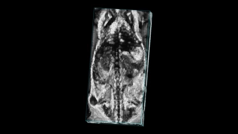
Mouse whole body with tumor
Whole body image of a mouse with a subcutaneous tumor visible on the flank, imaged with high frequency ultrasound on the Vevo F2 system.
ICG localization in tumor

Oxygen saturation in tumor
Publications
TOP PAPER
Assessment of Transarterial Chemoembolization Using Super-resolution Ultrasound Imaging and a Rat Model of Hepatocellular Carcinoma
Ultrasound in Medicine & Biology
,
TOP PAPER
Early diagnosis of bladder cancer by photoacoustic imaging of tumor-targeted gold nanorods
Photoacoustics
,
TOP PAPER
Magnetic black phosphorus microbubbles for targeted tumor theranostics
Nanophotonics
,
TOP PAPER
Morphological, functional, and molecular assessment of breast cancer bone metastases by experimental ultrasound techniques compared with magnetic resonance imaging and histological analysis
Bone
,
TOP PAPER
Contrast-Enhanced Multispectral Photoacoustic Imaging for Irregular Hepatectomy Navigation: A Pilot Study
ACS Biomaterials Science & Engineering
,
TOP PAPER
Tetrazine-Derived Near-Infrared Dye as a Facile Reagent for Developing Targeted Photoacoustic Imaging Agents
Molecular Pharmaceutics
,
TOP PAPER
Quantification of Murine Pancreatic Tumors by High-Resolution Ultrasound
Methods in Molecular Biology
,
TOP PAPER
Spectral Signatures in the Different Layers of the Human Eyelid by Photoacoustic Imaging
Lasers in Surgery and Medicine
, Request a Quote or Demo