This recent study by Berritto et al. highlights the benefits of using Ultra High Frequency (UHF) Ultrasound to visualize anatomical detail of the nail; and represents the first article published using the Vevo MD.
Article Summary:
Nail disease currently diagnosed by clinical exam since patients want to avoid biopsy
New methods of non-invasively diagnosing nail pathologies is desired
Study represents the first anatomical description of the healthy nail with UHF ultrasound
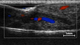
High resolution allows visualization of: layers of the nail plate, proximal and lateral nail folds, nail matrix and nail bed
Color Doppler can be used for straight capillaries and interlaced arterials and venules that supply and drain the nail
Can visualize anatomical detail unable to be seen with conventional ultrasound
Conclusion:
The Vevo MD with Ultra High Frequency Ultrasound technology is an effective tool at imaging nail bed anatomy and other dermatological applications. The high resolution can be used to assist in diagnosing nail pathologies and in preoperative planning.
1. Berritto, D. et al. Ultra high-frequency ultrasound: New capabilities for nail anatomy exploration. J. Dermatol. 1–4 (2016). doi:10.1111/1346-8138.13495