
This recent study by Scheepbouwer et al, highlights the benefits of a multimodal imaging approach by using Bioluminescence (BLI), High Resolution Ultrasound (HRUS) and Photoacoustics (PAI) to image orthotopic tumor proliferation over a 30 day time period.
The complementary abilities of these modalities allows detection of tumour heterogeneity and accurate quantification of tumour growth by providing anatomical, functional and molecular information.
Article Summary:
- Luciferase-expressing MB49 cells were implanted orthotopically into the murine bladder wall
- HRUS and PAI (with the Vevo LAZR system) as well as BLI was employed at regular time intervals over a four week time period
- At the end of experiment, tumours were excised and analysed by histopathology and immuno-histochemistry (IHC)
- Early-stage tumours were detected by HRUS, before BLI showed a luminescence signal
- Tumour-bearing mice showed an increased BLI signal over time and a slight decrease between days 17 and 24 whereas HRUS still detected tumour progression
- PAI oxygenation measurements showed that early-stage tumours were highly oxygenated
- With increased tumour volumes, average oxygen saturation levels dropped and hypoxic regions in the tumour were identified.
- Detected a 40% decrease in tumor oxygenation in treated vs. control mice
Conclusion:
BLI can visualize tumour cell proliferation non-invasively with high sensitivity and can differentiate tumour tissue from infection more easily than HRUS. However, BLI imaging tends to lose the spatial resolution while imaging orthotopic tumours. The BLI signal intensity is heavily dependent on oxygen/ATP and it may decrease even though the tumour is still growing, due to a possible hypoxic core. PAI with HRUS has the advantage of obtaining functional and molecular information in 3D, leading to better understanding of tumour development, and monitoring of therapeutic effects. All together, the combination of BLI, HRUS and PAI provides an attractive platform for monitoring bladder tumours longitudinally and for better prognosis of tumour proliferation and heterogeneity.
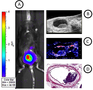
Comparison of BLI, HRUS and PAI images with macroscopic analysis of tumor volumes. (A) BLI image, (B) longitudinal section of the bladder with HRUS, (C) PAI image and (D) ex vivo bladder Histo-pathological image for comparison of multimodal images with macroscopic analysis of tumor volumes.
Reference:
Scheepbouwer C, Meyer S, Burggraaf MJ, Jose J, Molthoff CFM, ‘A Multimodal Imaging Approach for Longitudinal Evaluation of Bladder Tumor Development in an Orthotopic Murine Model’. PLoS ONE 11(8): e0161284, (2016). doi: 10.1371/journal.pone.0161284