Featured Images
Browse Research Ultrasound Images
High-resolution ultrasound from FUJIFILM VisualSonics allows you to visualize blood flow in an insect as small as a fruit fly (Drosophila) shown below. This video was captured using the MX700 probe on the Vevo 3100 ultrasound system in Color Doppler mode. Image courtesy of Thomas Jefferson University.
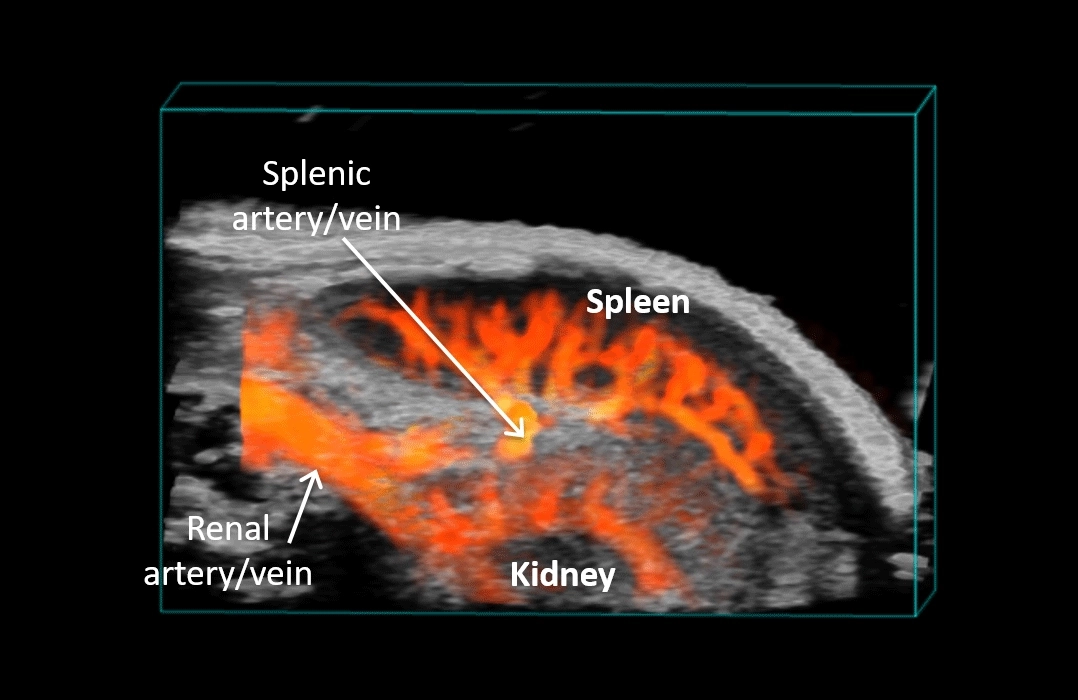
Detailed vasculature in the murine spleen and kidney visualized in 3D using the Vevo F2
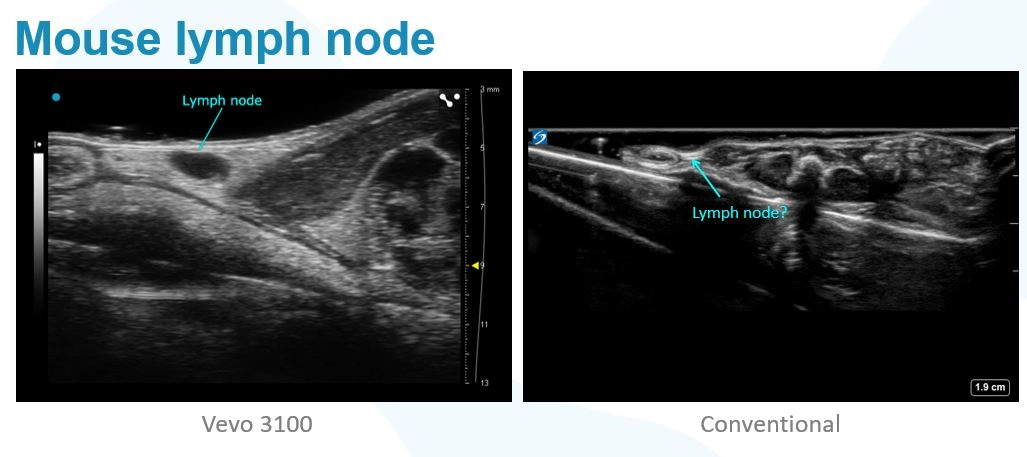
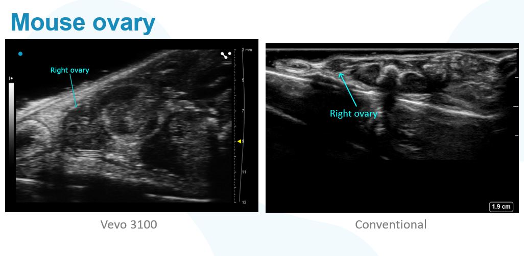
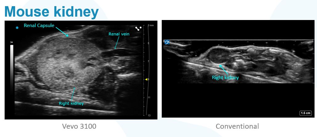
Shown here are some views of mouse anatomy using conventional ultrasound technology side by side ultra high frequency ultrasound images acquired using our Vevo 3100 system. Seeing more matters in research.
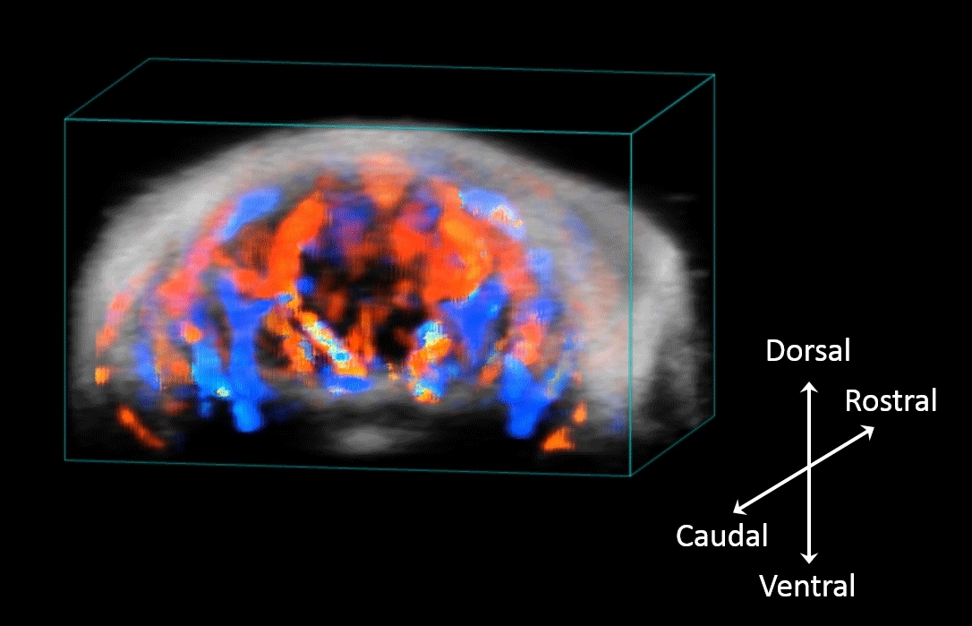
3D rendered and coregistered ultrasound (greyscale) and color Doppler (red and blue) image of the mouse brain. Color Doppler showing cerebral vasculature, highlighting major arteries and vein. Images were acquired non-invasively through intact skull and scalp.
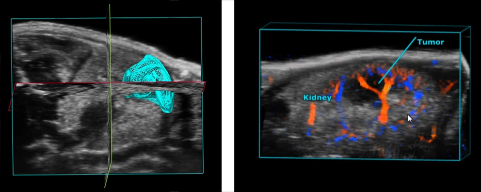
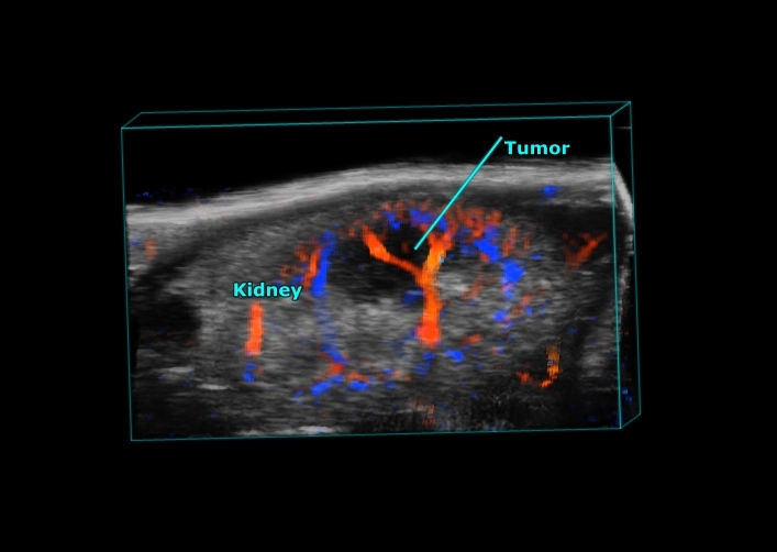
Kidney tumor in the mouse. Imaged in 3D with the tumor traced in blue (left) and imaged with Color Doppler in 3D showing vasculature (right).
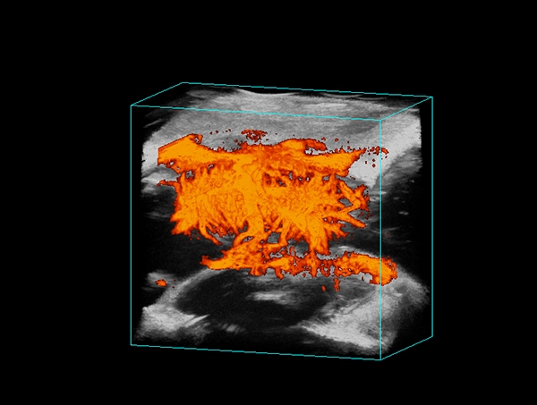
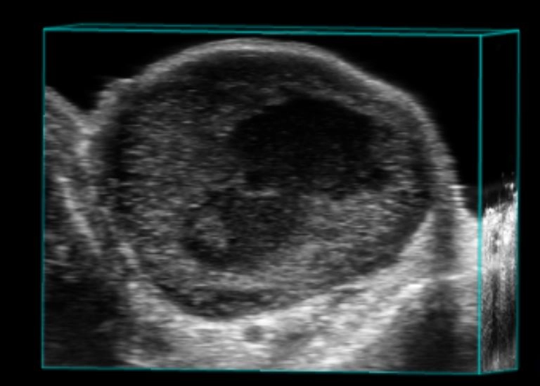
3D rendered and coregistered ultrasound (greyscale) and power Doppler (orange) image of a mouse brain. Images were acquired through a cranial window, looking at blood flow, showing the dense vasculature of the brain.