Fibrosis
Detect in vivo Fibrosis in Preclinical Models
Shear Wave Elastography all new and fully integrated on the Vevo F2!
.")
Shear wave elastography induces shear waves via ultrasound push pulse(s).
Why Image Fibrosis?
As tissue becomes damaged during disease progression, added collagen deposits begin to result in stiffer tissue. This occurs in numerous tissue types across the body. Most notably, as liver disease progresses towards liver cirrhosis, the increased collagen content results in increased fibrosis and stiffness in the liver. Elastography is one of the non-invasive ways clinicians use imaging to assess and visualize the level of fibrosis in the liver and the extent of disease progression.
Preclinically, assessing fibrosis non-invasively in small animal models of disease is an important step when studying liver disease or other disease models where fibrosis is common.
What is Elastography?
Shear Wave Elastography is a non-invasive imaging tool used to detect tissue stiffness. Low-frequency vibrations, or shear waves, are introduced into the tissue, then detected using ultrasound. Shear wave speed is used as a measure of tissue stiffness; faster shear wave propagation equates to a stiffer tissue.
How Preclinical Elastography Can Advance Your Research
- Visualize tissue stiffness non-invasively
- Easily quantify stiffness and shear wave velocity
- Assess liver fibrosis with high anatomical resolution
Request a Quote or Demo
Elastography in Action
Shear Wave Elastography Mode
Image Gallery

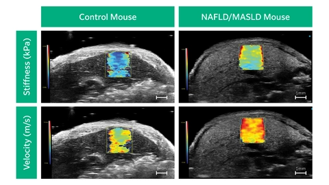
Shear Wave Elastography: NAFLD/MASLD Mouse - Velocity
Liver in a NAFLD/MASLD Mouse showing Velocity (m/s) through Shear Wave Elastography scanned using a UHF29x transducers on a Vevo F2 system.

Shear Wave Elastography: NAFLD/MASLD Mouse - Stiffness
Liver in a NAFLD/MASLD Mouse showing Stiffness (kPa) through Shear Wave Elastography scanned using a UHF29x transducers on a Vevo F2 system.

Shear Wave Elastography: Control Mouse - Velocity
Liver in a Control Mouse showing Velocity (m/s) through Shear Wave Elastography scanned using a UHF29x transducers on a Vevo F2 system.

Shear Wave Elastography: Control Mouse - Stiffness
Liver in a Control Mouse showing Stiffness (kPa) through Shear Wave Elastography scanned using a UHF29x transducers on a Vevo F2 system.
