
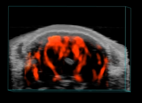
Mouse Brain Vasculature
3D Power Doppler rendering of a mouse brain, highlighting cerebral vessels.
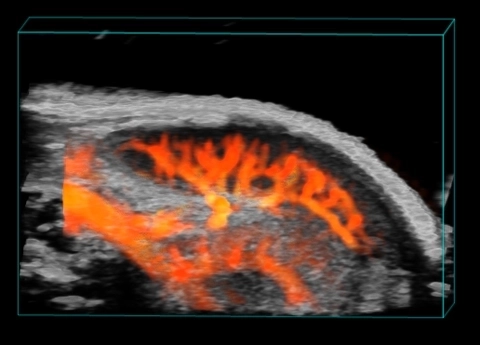
3D Spleen and Kidney Vasculature
3D Power Doppler rendering of a mouse spleen and kidney, highlighting splenic and renal vasculature.

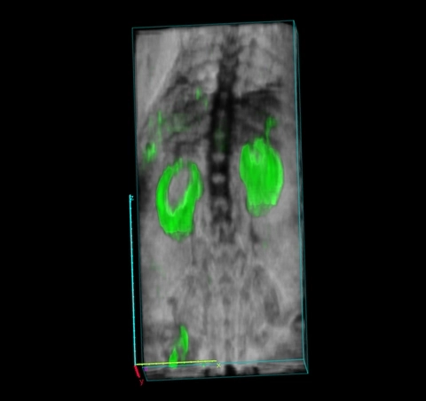
Biodistribution of photoacoustic contrast agent and signals from oxy and deoxyhemoglobin
Whole body biodistribution of photoacoustic contrast agent Angiostamp800TM (green), along with signals from oxy (red) and deoxyhemoglobin (blue).
Whole body biodistribution
Whole body biodistribution of photoacoustic contrast agent Angiostamp800TM (green)
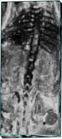
Whole Body Image of Mouse
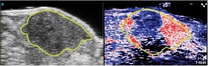
Pancreatic tumor oxygen saturation
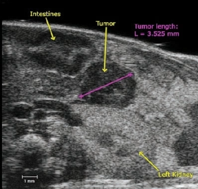
Pancreatic tumor size
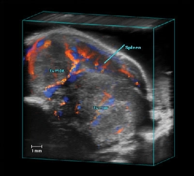
Pancreatic Tumor Color Doppler 3D
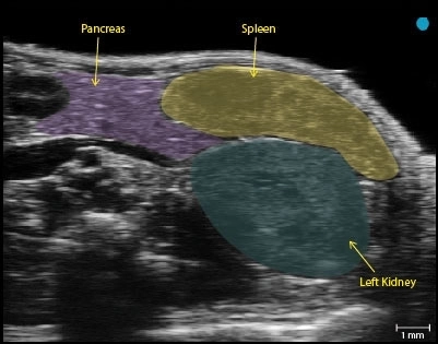
Healthy Pancreas - Tail
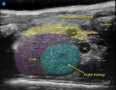
Healthy Pancreas - Head

Radial Artery - Adult Female

Palm - Adult Female
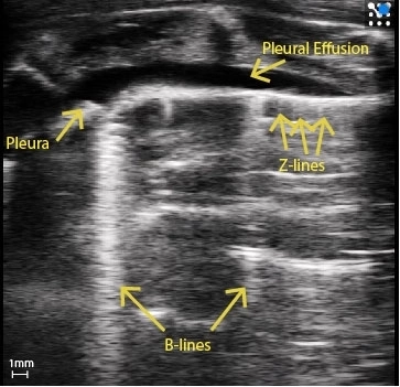
Rat Pleural Effusion
Rat with pleural effusion and pleural thickening. Clear B-Lines can be seen as well as Z-Lines.
Image courtesy of : M.Sc. Niklas Hegemann, Kübler lab, Institute of Physiology, Charité-Universitätsmedizin Berlin & Dr. Jana Grune, Nahrendorf lab, Center for Systems Biology, Massachusetts General Hospital.
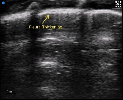
Pleural thickening
Substantial pleural thickening in the rat lung.
Image courtesy of : M.Sc. Niklas Hegemann, Kübler lab, Institute of Physiology, Charité-Universitätsmedizin Berlin & Dr. Jana Grune, Nahrendorf lab, Center for Systems Biology, Massachusetts General Hospital.
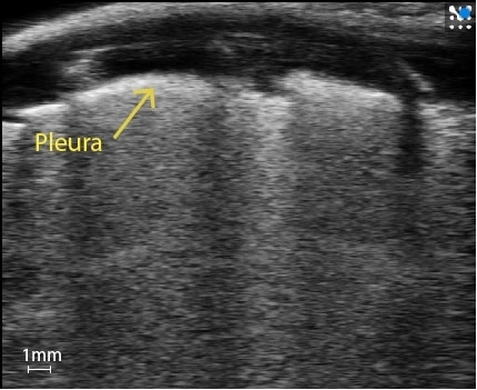
Murine White Lung
Mouse lung covered in B-Lines, creating white lung.
Image courtesy of : M.Sc. Niklas Hegemann, Kübler lab, Institute of Physiology, Charité-Universitätsmedizin Berlin & Dr. Jana Grune, Nahrendorf lab, Center for Systems Biology, Massachusetts General Hospital.