Submitted by Tatjana Opacic, Experimental Molecular Imaging.
Super-resolution contrast-enhanced ultrasound (CEUS) methods have been introduced to visualize and quantify microvasculature beyond the resolution limit of the ultrasound devices. In this study, we developed a new method called motion model ultrasound localization microscopy (mULM) for morphological and functional characterization of tumors at super-resolution. In tumor-bearing mice, we showed that super-resolution ultrasound imaging is feasible with Vevo ultrasound technology, standard frame rates and short measurement times.
We evaluated mULM in three tumor xenografts with different vascular pattern (A431, MLS and A549). Data were acquired with a Vevo 2100 system (FUJIFILM VisualSonics, Toronto, ON, Canada) using the 50 MHz transducer after intravenous injection of hard-shell polybutylcyanoacrylate (PBCA) microbubbles (MB). mULM analysis, performed as described by Ackermann et al 1, provided information about the relative blood volume (rBV), blood flow velocity and blood flow direction, depicted in corresponding parameter maps at super-resolution.
Additionally, we extracted novel parameters, distances to the vessels with low and high velocity, which are capable to spatially and functionally characterize tumor vasculature.
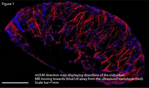
mULM precisely captured the vascular tumor architectures and was capable of distinguishing the different tumor types. In particular, textural parameters describing the distances to vessels of high or low velocities, which could only be obtained by mULM, showed supremacy to discriminate the tumor models. The velocities and directions of the individual microbubbles were displayed in fine detail in the color-coded velocity and direction maps. In this context, direction maps could be generated by coding MB moving towards and away from the transducer in blue and red, respectively, which would correspond to conventional Doppler imaging (Figure 1).
In summary, using the Vevo 2100 imaging system from FUJIFILM VisualSonics, Inc. we established mULM as a novel super resolution US analysis method. The multiple parameters deriving from mULM analysis can be used for US-based radiomics and provide important insights into tumor vascularization. We strongly believe that this method will advance preclinical tumor research and clinically help to improve tumor diagnosis and treatment monitoring.
For more information on our work, please see Opacic T et al. Nat Commun. 2018 Apr 18;9(1):1527. doi: 10.1038/s41467-018-03973-8.
References:
1. Ackermann, D. & Schmitz, G. Detection and Tracking of Multiple Microbubbles in Ultrasound B-Mode Images. IEEE Trans Ultrason Ferroelectr Freq Control 63, 72-82 (2016).