Submitted by Yu-Qing Zhou, PhD.
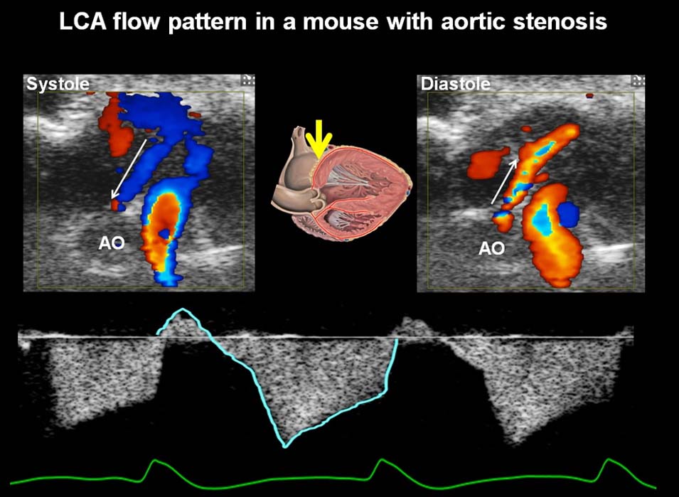
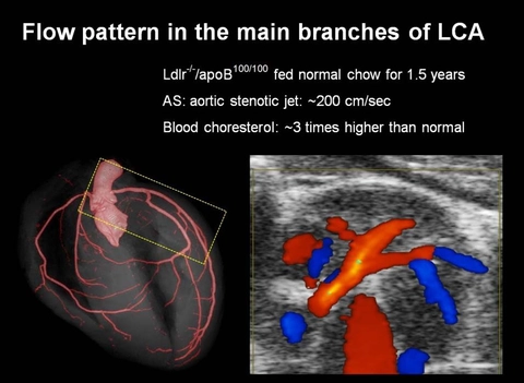
At the Mouse Imaging Centre of the Hospital for Sick Children in Toronto, we have collaborated with VisualSonics Inc. since early 2000 and have used their products from the very first generation to the most advanced versions. With the state-of-the-art technologies from VisualSonics, we have conducted very extensive research on cardiovascular physiology and in vivo phenotyping of genetic mouse models of human diseases. We have established comprehensive methodologies for mouse cardiovascular imaging using high frequency ultrasound (Physiological Genomics 18:232-244;2004), and successfully applied the established methodology to the phenotyping of mutant mouse models with human diseases. These methods have been widely accepted in this field. In recent years we have used the Vevo2100 imaging system together with other micro-imaging technologies to evaluate the association between aortic flow dynamics and atherogenesis in mouse models and also to study the mouse fetal and placental physiology (Physiological Genomics 46:602-614;2014). Vevo systems have played a central role in more than 30 publications from our group and continue to be one of our most heavily used imaging technologies.