
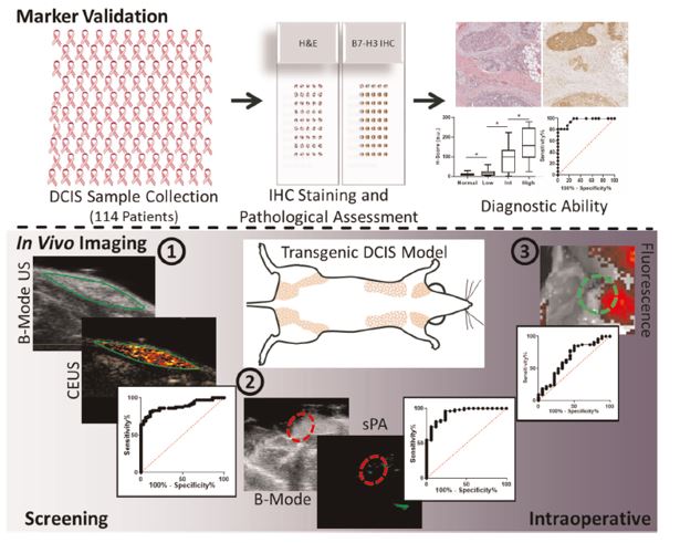
In this recent study by Bachawal et al., authors used the multimodal imaging capabilities of the Vevo imaging system to evaluate the aggressiveness of ductal carcinoma in situ (DCIS) through B7-H3 targeted ultrasound and photoacoustic molecular imaging.
Article Summary:
- DCIS accounts for 20% of the breast cancer detected through screening, with surgical resection being the mainstay treatment.
- DCIS is a pre-invasive lesion that may not progress to invasive carcinoma.
- There is a need for non-invasive methods of monitoring disease progression so that patient over treatment can be reduced, along with healthcare costs.
- Increased expression of B7-H3 was shown through immunohistochemical staining to be associated with more invasive grades of DCIS, making it an ideal biomarker for the monitoring of disease progression.
- A transgenic mouse model of breast cancer development was used for this study.
- Molecule ultrasound (US) imaging using targeted microbubbles was used to determine normal vs DCIS tissue for screening purposes.
- Photoacoustic and fluorescence imaging using anti-B7-H3 antibody conjugated ICG dye was used to detect DCIS margins intraoperatively.
- Contrast ultrasound differentiated DCIS from normal tissue with an AUC of 0.89.
- Photoacoustic (PA) imaging with targeted contrast agent showed high specificity and was able to differentiate small foci of DCIS (< 1mm) from normal tissue.
Conclusion:
In this study the authors showed that the expression level of B7-H3 is correlated with the nuclear grade of DCIS. Used in combination with appropriate contrast agents, targeted B7-H3 imaging with US, PA and fluorescence was able to sensitively detect DCIS. Posing strategies for potential clinical use.
- Bachawal, S., Bean, G. R., Krings, G. & Wilson, K. E. Evaluation of ductal carcinoma in situ grade via triple-modal molecular imaging of B7-H3 expression. npj Breast Cancer 6, 14 (2020).