Bacterial infection is a major cause of morbidity and mortality, especially in the case of antibiotic-resistant strains of S. aureus. A small animal model that uses noninvasive imaging provides a valuable system to study pathogenesis, its treatments, and diagnostic approaches.
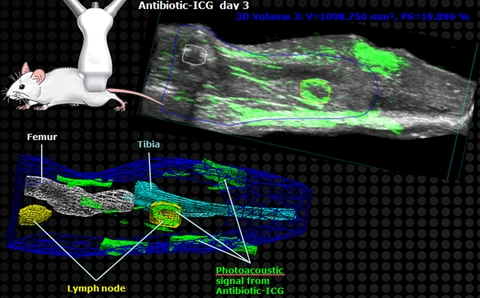
In collaboration with Lloyd Miller, Associate Professor in the Department of Dermatology at Johns Hopkins University, we used micro-ultrasound and photoacoustic imaging with the Vevo LAZR system to investigate such a model.
An antibiotic tagged with an optical dye was administered to animals that had a subdermal S. aureus infection in the hindlimb and co-registered ultrasound and multispectral photoacoustic images were acquired in 3D (ultrasound/photoacoustic image). Using the VevoLAB software, 3D regions of interest were drawn on the images to segment out various tissues such as bones and lymph nodes as well as photoacoustic signal arising from the optical dye, and these ROIs were displayed as wireframe renderings (wireframe image). In this way, precise anatomical localization and quantification of the signal can be performed, thus yielding data about the extent and location of the bacterial infection.