Oxy-Hemo Mode
Assess Hypoxia Within the Tumor Microenvironment and More
Take advantage of having oximetry data fused with high resolution anatomical images.
While methods exist for measuring blood oxygenation, they can be invasive, low temporal and/or spatial resolution, superficial or expensive. Because oxy- and deoxyhemoglobin absorb light differently at different wavelengths, photoacoustic imaging can be used to generate a high resolution parametric map of the oxygen saturation of blood completely non-invasively. This allows for the real-time monitoring of functional processes with the added advantage of having this oximetry data fused with high resolution anatomical images.
PA EKV stands for Photoacoustic ECG-gated Kilohertz Visualization. This unique software allows for photoacoustic imaging with increased temporal resolution and is ideal for cardiovascular applications. Learn more.
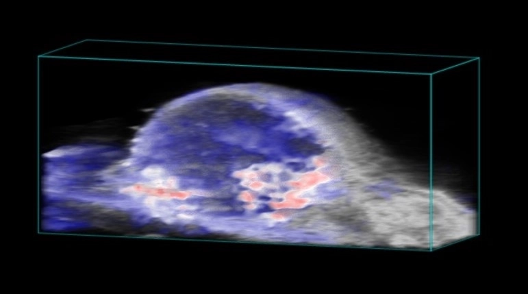
Subcutaneous tumor showing spatial distribution of oxy and deoxyhemoglobin for hypoxia assessment.
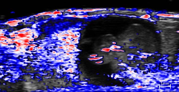
E14 embryo (right) and associated placenta (left) showing oxygenation distribution.