Submitted by Maria Stansczak, MS, Co-Authors: Jing Chen-Roetling, John Eisenbrey, Flemming Forsberg, Ji-Bin Liu, Raymond Regan - Departments of Radiology and Emergency Medicine, Thomas Jefferson University, Philadelphia
Intracerebral hemorrhages (ICH) is a cause of stroke for which no effective drug therapy exists. Our work investigates a novel mouse model of ICH with high frequency ultrasound (US) and photoacoustics for long-term screening of potential preclinical therapeutics.
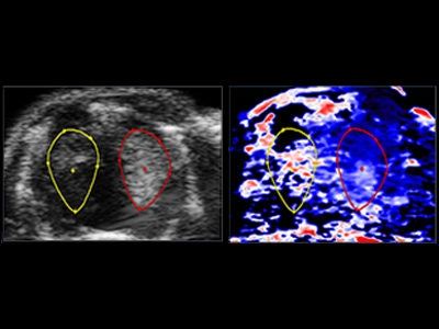
To develop the ICH model, mice underwent right partial craniotomies followed by unilateral direct intracerebral injections of collagenase material. High frequency (40-70 MHz) bilateral US and photoacoustic imaging was performed 1,2 and 8 days post injection with the Vevo 2100/LAZR. ICH characteristics including size, echogenicity, vascular perfusion status, and saturated oxygen levels were measured and compared to the contralateral cerebral hemisphere.
The size of the hemorrhage ranged from 1.9 to 3.6 mm, with smaller sizes measured at 8 days post injection. Initial homogeneous hyperechogenicities was visualized within the affected areas 24 hours post injection. Hypoechogenic areas developed within the surrounding hyperechogenicities 48 hours post injection and more prominenetly visualized 8 days post injection. Power Doppler of the affected side revealed decreased vascular perfusion patterns 24 and 48 hours post injection. A return to symmetrical vascular perfusion patterns was observed 8 days post injection. Photoacoustic imaging of the affected versus unaffected sides showed decreased oxygen levels 24 and 48 hours post injection (by 48% and 14%, respectively), before equalizing on day 8.
We concluded that assessment of a murine model of ICH can be achieved with high frequency US and photoacoustic imaging. Changes in ICH size, echogenicity, vascular perfusion and oxygen levels can be observed 24 hours, 48 hours and 8 days post injury. This could be a useful longitudinal imaging tool for characterizing ICH in a murine model, and should speed translation of experimental therapeutics.
Maria Stanczak is an ultrasound researcher and faculty member at Thomas Jefferson University in Philadelphia, PA. Her research interests include ultrasound contrast agents, subharmonic imaging, high frequency and photoacoustic scanning. Her recent award-winning research presented at RSNA 2015 focused on creation and monitoring of a novel mouse model for intracerebral hemorrhage using Vevo ultrasound and photoacoustics.