Submitted by Terri A. Swanson, Senior Scientist, Comparative Medicine, Pfizer Inc. and Theresa Tuthill, Head of Cardiovascular, Metabolic, and Musculoskeletal Imaging for the Clinical and Translational Imaging group at Pfizer Inc.
Terri Swanson, Theresa Tuthill and their team at Pfizer share their story and navigate through some of the translational challenges in developing imaging methods for fibrosis. While fibrosis (tissue scarring) is a normal response to injury, the deposition of connective tissue to form scars challenges the organ to function normally. The mechanism of fibrosis is still unclear, and its onset/progression is generally visualized via biopsy and histology. Drug development for fibrotic diseases brings a more demanding challenge – characterizing and monitoring the onset and progression of fibrosis for in vivo longitudinal studies to assist in Pfizer’s efforts to develop novel treatments for these diseases.
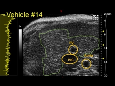
This challenge demanded the knowledge to generate the right in vivo model for longitudinal monitoring by high frequency ultrasound waves. Pfizer teams led by Terri Swanson and Theresa Tuthill explored a variety of techniques and quantitative analyses to address this complex challenge. In a recent webinar, they share how they brought their individual expertise in high frequency ultrasound to address the science of developing therapeutics for fibrosis in the liver and heart.