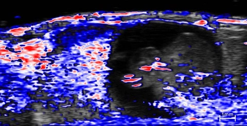
In vivo co-registered high-resolution ultrasound (greyscale) and photoacoustic (red, white and blue) image of an E14 embryo and its associated placenta (on left of each image). The photoacoustic signal represents a parametric map of oxygen saturation with red values representing higher sO2. The image was acquired while the mother was breathing medical air (20% O2). Oxygen saturation can be distinguished within differing placental and embryonic anatomy.