Vevo MD Gallery
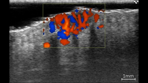
Color Doppler in Basal Cell Carcinoma Lesion
Color Doppler showing blood flow within a basal cell carcinoma lesion in a patient using ultra high frequency ultrasound on the Vevo MD.
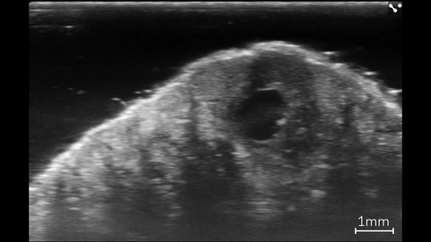
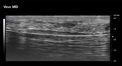
Basal Cell Carcinoma Lesion
High resolution image of a basal cell carcinoma lesion in a patient, imaged with the Vevo MD.
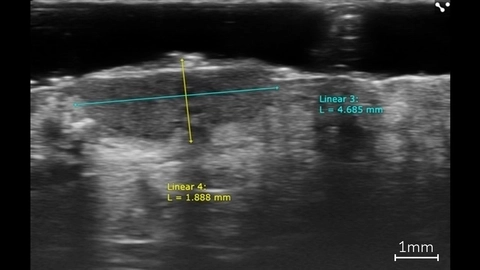
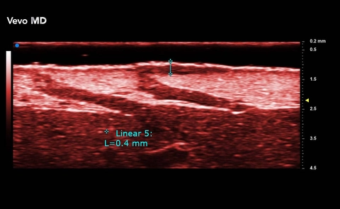
Linear Measurements of a Basal Cell Carcinoma
Linear measurements of a basal cell carcinoma in a patient using ultra high frequency ultrasound on the Vevo MD.
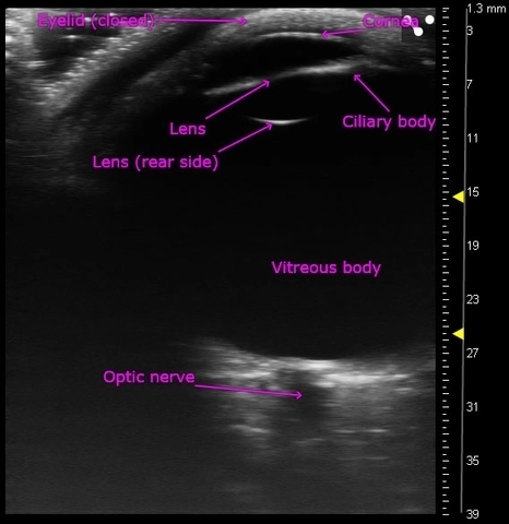
Entire adult human eye images with ultra high frequency ultrasound
Acquired using the Vevo MD.
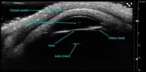
Anterior chamber of the adult human eye
Acquired using the Vevo MD.

Radial Artery - Adult Female

Palm - Adult Female
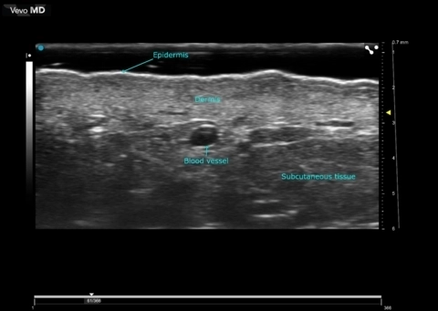
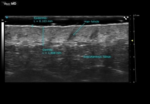
High Resolution Ultrasound Dermal Imaging
Scanned using a UHF70 transducer on the Vevo MD.
Facial Skin
Ephelis
Ephelis
A1 Pulley
A1 Pulley
