MSK Gallery
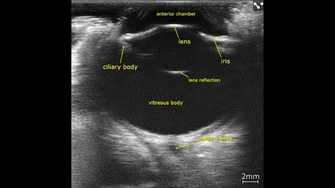
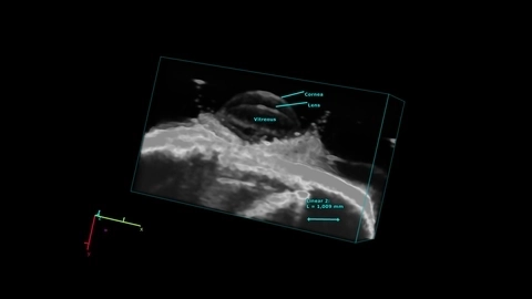
Canine eye in vivo
Entire canine eye visualized in vivo with the Vevo F2, scanned using a UHF22x transducer.
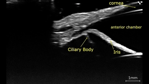
Canine eye angle of the anterior chamber
Canine eye imaged in vivo on the Vevo F2 scanned using the UHF71x.
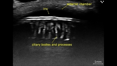
Canine eye, Ciliary body and processes
In vivo canine eye with ciliary processes, imaged with high-frequency on the Vevo F2 scanned using the UHF71x.
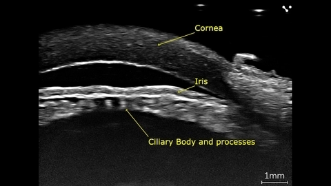
Porcine eye, Ciliary body and processes
Porcine eye imaged ex-vivo using the Vevo F2 with a UHF71x transducer.
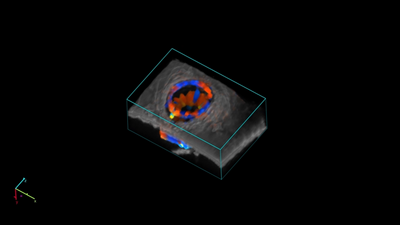
Mouse Eye in 3D
Mouse eye scanned using the UHF71x transducer using the Vevo F2 in 3D.


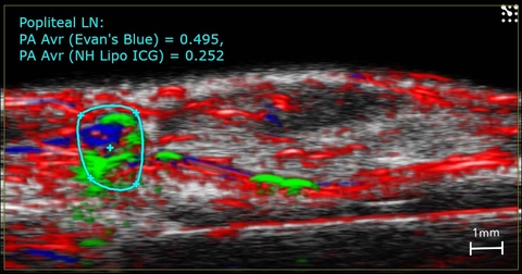
Imaging of lymphatics in the mouse.
High-resolution ultrasound (greyscale) and photoacoustic (red, blue and green) image of the mouse hindlimb showing Evan's Blue (blue) and ICG (green) dye in the lymphatic vessels and the popliteal lymph node.
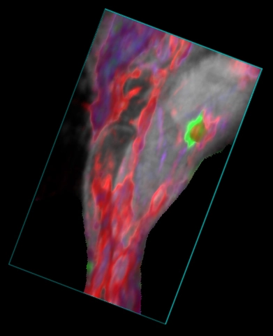
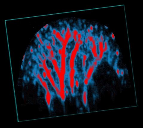
Mouse hindlimb showing blood and ICG
3D rendered high-resolution ultrasound (greyscale) and spectrally unmixed photoacoustic (red, blue and green) image of the mouse hindlbimb. The green color indicates ICG dye in the popliteal lymph node after a hindpaw injection, the red color indicated oxygenated hemoglobin and the blue color indicates deoxygenated hemoglobin.
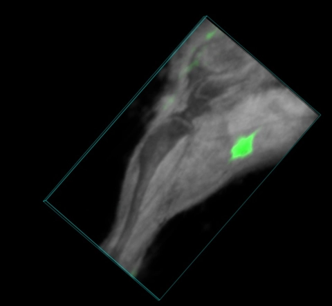
Mouse hindlimb with ICG in lymph node
3D rendered high-resolution ultrasound (greyscale) and spectrally unmixed photoacoustic (green) image of the mouse hindlbimb. The green color indicates ICG dye in the popliteal lymph node after a hindpaw injection.
Mouse ear vasculature
3D rendering of a photoacoustic parametric map of oxygen saturation in the mouse ear. Red pixels indicate higher sO2 values.
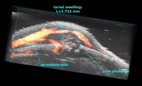
Arthritic Mouse Paw
Rendered 3D power Doppler image of an arthritic mouse paw with bone erosion.
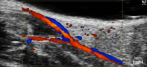
Femoral Artery Bifurcation
Bifurcation of the femoral artery in the mouse seen using color Doppler.
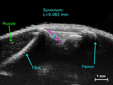
Synovium in a Healthy Knee
Synovium in a healthy mouse knee.
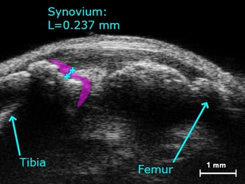
Synovium in Collagen-induced Arthritic Knee
Thickened synovium in the mouse knee due to collagen-induced arthritis.