Colorectal Gallery

Orthotopic Pancreatic Tumor
High-resolution ultrasound image of an orthotopic pancreatic tumor outlining anatomy.
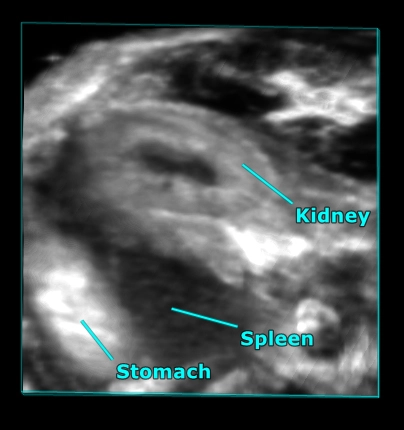
Mouse abdominal organs
3D rendered high-resolution ultrasound image of the mouse abdomen showing the kidney, stomach and spleen.
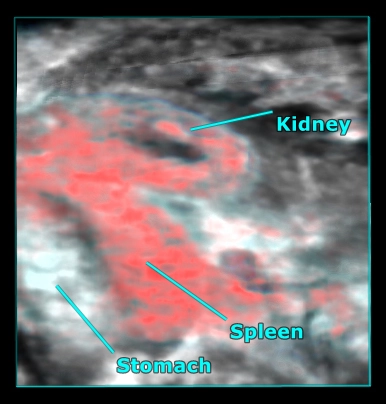
Mouse abdominal organs
3D rendered high-resolution ultrasound (greyscale) and photoacoustic (red) image of the mouse abdomen showing oxygen saturation in the kidney and spleen.
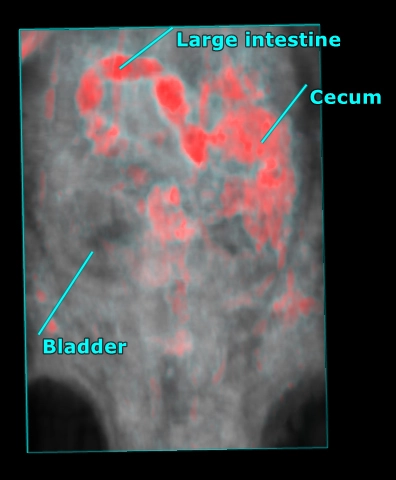
Mouse abdomen
3D rendered high-resolution ultrasound (grey) and photoacoustic (red) image of the whole mouse abdomen showing highly absorbing gut contents overlaid with anatomy as visualized with ultrasound.
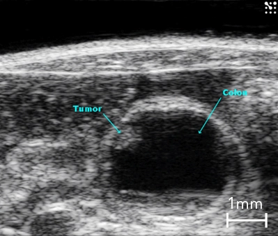
Colon Cancer
B-Mode image of the mouse colon in cross-section with presence of a tumor highlighted.
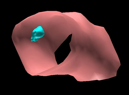
3D Surface Volume of the Mouse Colon with Tumor
Surface view of a 3D volume of a section of mouse colon (in pink) with a tumor (in blue).
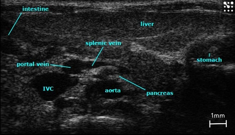
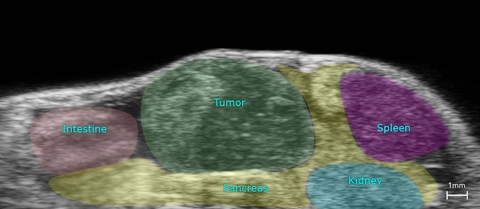
Major Structures in the Mouse Abdomen
B-Mode image of the mouse abdomen with all major structures labelled.