Oncology Gallery
Breast tumor in 3D
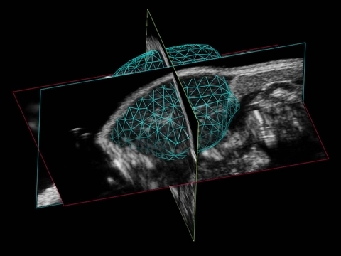
Tumor 3D
3D Volume Reconstruction of a Murine Tumor
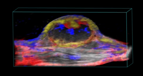
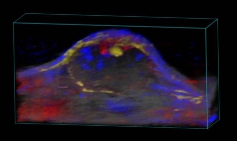
Nanoparticle distribution in tumor
3D rendered high-resolution ultrasound (greyscale) and spectrally unmixed photoacoustic (red, blue and gold) image of a subcutaneous tumor showing nanoparticle distribution (yellow) as well as oxygenated (red) and deoxygenated (blue) hemoglobin signal.
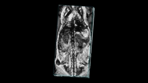
Mouse whole body with tumor
Whole body image of a mouse with a subcutaneous tumor visible on the flank, imaged with high frequency ultrasound on the Vevo F2 system.
ICG localization in tumor

Oxygen saturation in tumor
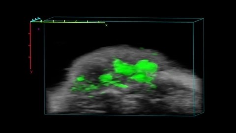
Perfusion in Tumor
3D rendered high-resolution ultrasound (greyscale) and nonlinear contrast (beige) image of a subcutaneous tumor showing perfusion in the tumor tissue.
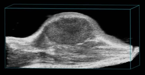
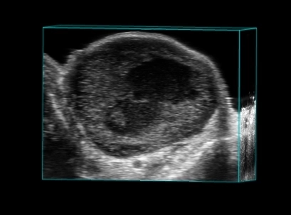
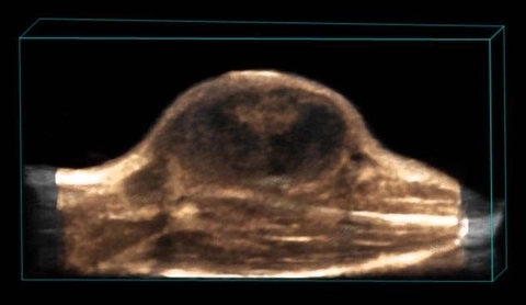
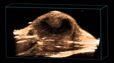
Subcutaneous tumor
3D rendered high-resolution ultrasound image of a subcutaneous tumor.
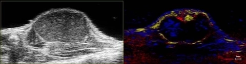
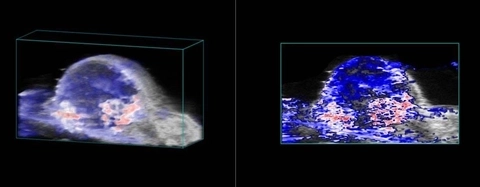
Nanoparticle distribution in tumor
High-resolution ultrasound (left) and spectrally unmixed photoacoustic (right) image of a subcutaneous tumor showing nanoparticle distribution (yellow) as well as oxygenated (red) and deoxygenated (blue) hemoglobin signal.
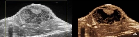
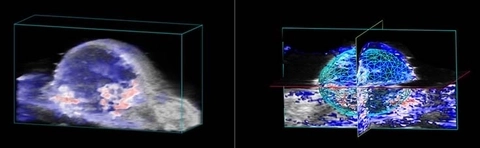
Perfusion in Tumor
High-resolution ultrasound (left) and nonlinear contrast (right) image of a subcutaneous tumor showing perfusion in the tumor tissue.
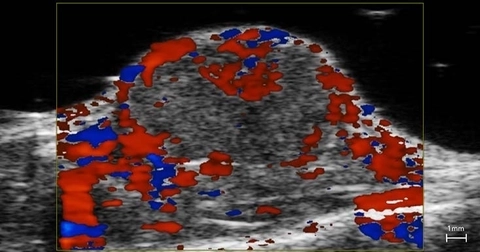
Blood flow in tumor
Perfusion in Tumor
3D rendered high-resolution nonlinear contrast image of a subcutaneous tumor showing perfusion in the tumor tissue.
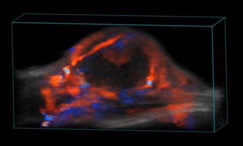
Blood flow in tumor
3D rendered high-resolution ultrasound (greyscale) and color Doppler (orange and blue) image of a subcutaneous tumor showing blood flow.
Nanoparticle distribution in tumor
3D rendered high-resolution ultrasound (greyscale) and spectrally unmixed photoacoustic (red, blue and gold) image of a subcutaneous tumor showing nanoparticle distribution (yellow) as well as oxygenated (red) and deoxygenated (blue) hemoglobin signal.
Oxygen saturation in tumor