Kidney Gallery
![Play](/themes/custom/ergo/assets/images/play.png)
Right Kidney
![Play](/themes/custom/ergo/assets/images/play.png)
Kidney with Shadow
![Play](/themes/custom/ergo/assets/images/play.png)
PW Doppler of Renal Artery Flow
Renal Artery Flow acquired through PW Doppler, scanned using a UHF57x transducer on the Vevo F2.
![B-mode of the ovary, spleen and kidney](/sites/default/files/styles/crop_thumbnail/public/2022-09/F2_UHF57_Mouse%20Ovary%20spleen%20kidney.jpg?itok=RhRcz3AM)
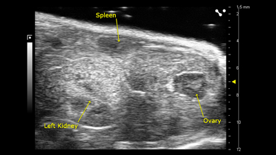
Ovary, spleen and kidney
![B-mode of the ovary, spleen and kidney](/sites/default/files/styles/crop_thumbnail/public/2022-09/F2_UHF57_Mouse%20Ovary%20spleen%20kidney.jpg?itok=RhRcz3AM)
B-mode image of the ovary, spleen and kidney, acquired using a UHF57x transducer on the Vevo F2.
![Color Doppler of the sagittal view in the left kidney](/sites/default/files/styles/crop_thumbnail/public/2022-09/F2_Sagittal%20view%20left%20kidney%20color.jpg?itok=vFwXGa8f)
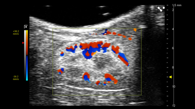
Sagittal View in the Left Kidney
![Color Doppler of the sagittal view in the left kidney](/sites/default/files/styles/crop_thumbnail/public/2022-09/F2_Sagittal%20view%20left%20kidney%20color.jpg?itok=vFwXGa8f)
Color Doppler image of the sagittal view in the left kidney, acquired using a UHF57x transducer on the Vevo F2.
![B-mode of intestine, right kidney, right lobe liver, portal vein, inferior vena cava, and adrenal gland](/sites/default/files/styles/crop_thumbnail/public/2022-09/F2_Right%20liver%20lobe%20adrenal%20gland.jpg?itok=f016Gdvw)
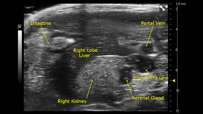
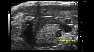
Right Liver Lobe and Adrenal Gland
![B-mode of intestine, right kidney, right lobe liver, portal vein, inferior vena cava, and adrenal gland](/sites/default/files/styles/crop_thumbnail/public/2022-09/F2_Right%20liver%20lobe%20adrenal%20gland.jpg?itok=f016Gdvw)
B-mode image of the right liver lobe and adrenal gland, acquired using a UHF57x transducer on the Vevo F2.
![B-mode of the right kidney, portal vein, and inferior vena cava](/sites/default/files/styles/crop_thumbnail/public/2022-09/F2_Right%20kidney%20portal%20IVC.jpg?itok=FtswyZX3)
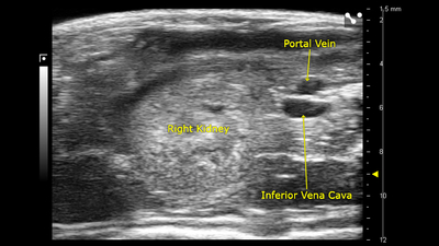
Right Kidney Portal System
![B-mode of the right kidney, portal vein, and inferior vena cava](/sites/default/files/styles/crop_thumbnail/public/2022-09/F2_Right%20kidney%20portal%20IVC.jpg?itok=FtswyZX3)
B-mode image of the right kidney, portal vein, inferior vena cava, acquired using a UHF57x transducer on the Vevo F2.
![B-mode of the right adrenal gland](/sites/default/files/styles/crop_thumbnail/public/2022-09/F2_Right%20adrenal%20gland.jpg?itok=aQgO4PES)
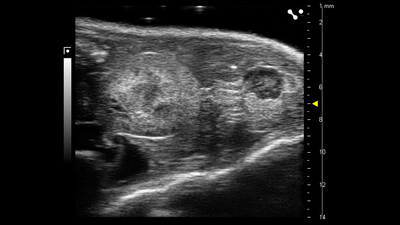
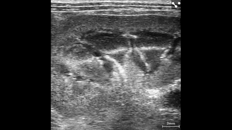
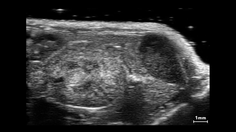
Right Adrenal Gland of the Mouse
![B-mode of the right adrenal gland](/sites/default/files/styles/crop_thumbnail/public/2022-09/F2_Right%20adrenal%20gland.jpg?itok=aQgO4PES)
B-mode image of the right adrenal gland of the mouse, acquired using a UHF57x transducer on the Vevo F2.
![PW Doppler measurements for renal artery flow](/sites/default/files/styles/crop_thumbnail/public/2022-09/F2_Renal%20artery%20flow%20measurements.jpg?itok=0luw52m3)
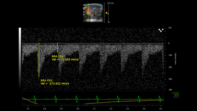
Renal Artery Flow Measurements
![PW Doppler measurements for renal artery flow](/sites/default/files/styles/crop_thumbnail/public/2022-09/F2_Renal%20artery%20flow%20measurements.jpg?itok=0luw52m3)
PW Doppler measurements for renal artery flow, acquired using a UHF57x transducer on the Vevo F2.
![B-mode of a mouse ovary and kidney](/sites/default/files/styles/crop_thumbnail/public/2022-09/F2_Mouse%20ovary%20and%20kidney%20UHF57x.jpg?itok=28gEjDzW)
Mouse ovary and kidney
![B-mode of a mouse ovary and kidney](/sites/default/files/styles/crop_thumbnail/public/2022-09/F2_Mouse%20ovary%20and%20kidney%20UHF57x.jpg?itok=28gEjDzW)
B-mode image of the mouse ovary and kidney, acquired using a UHF57x transducer on the Vevo F2.
![Color Doppler of renal vein, artery, left kidney and spleen](/sites/default/files/styles/crop_thumbnail/public/2022-09/F2_Kidney%20color%20%20spleen%20labeled.jpg?itok=9sRqmo_3)
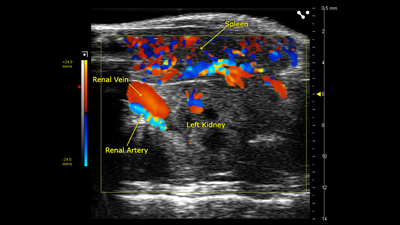
Color Doppler of Mouse Kidney
![Color Doppler of renal vein, artery, left kidney and spleen](/sites/default/files/styles/crop_thumbnail/public/2022-09/F2_Kidney%20color%20%20spleen%20labeled.jpg?itok=9sRqmo_3)
Color Doppler image of renal vein, artery, left kidney and spleen, acquired using a UHF57x transducer on the Vevo F2.
![B-mode of the sagittal kidney](/sites/default/files/styles/crop_thumbnail/public/2022-09/F2%20UHF57%20sagittal%20kidney.jpg?itok=_Z70u-0R)
Sagittal Kidney
![B-mode of the sagittal kidney](/sites/default/files/styles/crop_thumbnail/public/2022-09/F2%20UHF57%20sagittal%20kidney.jpg?itok=_Z70u-0R)
B-mode image of the sagittal kidney acquired using a UHF57x transducer on the Vevo F2.
![B-mode of the left kidney, spleen and pancreas](/sites/default/files/styles/crop_thumbnail/public/2022-09/F2%20UHF57%20L%20Kidney%20Spleen%20Pancreas.jpg?itok=X87h5OOS)
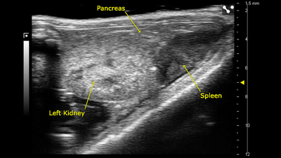
Left Kidney, Spleen and Pancreas
![B-mode of the left kidney, spleen and pancreas](/sites/default/files/styles/crop_thumbnail/public/2022-09/F2%20UHF57%20L%20Kidney%20Spleen%20Pancreas.jpg?itok=X87h5OOS)
B-mode image of the left kidney, spleen and pancreas acquired using a UHF57x transducer on the Vevo F2.
![Play](/themes/custom/ergo/assets/images/play.png)
Mouse kidney NLC
![Rat kidney vasculature in 3D color - UHF29x - 15-29MHz.jpg](/sites/default/files/styles/large/public/media-files/Rat%20kidney%20vasculature%20in%203D%20color%20-%20UHF29x%20-%2015-29MHz.jpg.webp?itok=uyQ9bjWg)
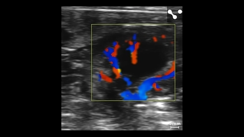
Color Doppler of Rat Kidney Vasculature
![Rat kidney vasculature in 3D color - UHF29x - 15-29MHz.jpg](/sites/default/files/styles/large/public/media-files/Rat%20kidney%20vasculature%20in%203D%20color%20-%20UHF29x%20-%2015-29MHz.jpg.webp?itok=uyQ9bjWg)
Color Doppler of rat kidney vasculature scanned using a UHF29x transducer.
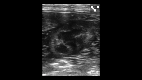
![Rabbit kidney - UHF29x.jpg](/sites/default/files/styles/large/public/media-files/Rabbit%20kidney%20-%20UHF29x.jpg.webp?itok=co0oOSnx)
Rabbit Kidney
![Rabbit kidney - UHF29x.jpg](/sites/default/files/styles/large/public/media-files/Rabbit%20kidney%20-%20UHF29x.jpg.webp?itok=co0oOSnx)
Rabbit kidney scanned using a UHF29x transducer.
![Murine renal vasculature in 3D.jpg](/sites/default/files/styles/large/public/media-files/Murine%20renal%20vasculature%20in%203D.jpg.webp?itok=LJDNBzi-)
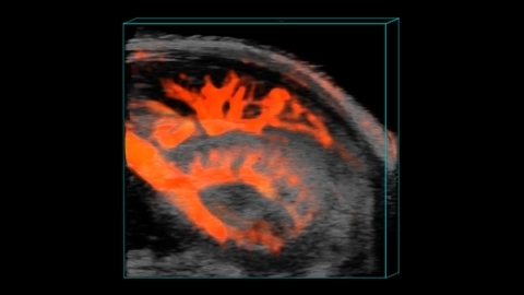
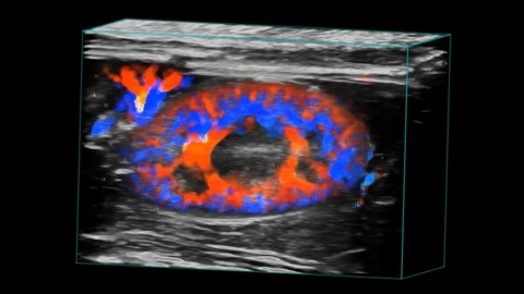
Murine Renal Vasculature in 3D
![Murine renal vasculature in 3D.jpg](/sites/default/files/styles/large/public/media-files/Murine%20renal%20vasculature%20in%203D.jpg.webp?itok=LJDNBzi-)
![Mouse kidney, spleen and pancreas.jpg](/sites/default/files/styles/large/public/media-files/Mouse%20kidney%2C%20spleen%20and%20pancreas.jpg.webp?itok=jkYP8cQP)
Mouse Kidney, Spleen and Pancreas
![Mouse kidney, spleen and pancreas.jpg](/sites/default/files/styles/large/public/media-files/Mouse%20kidney%2C%20spleen%20and%20pancreas.jpg.webp?itok=jkYP8cQP)
![Left kidney in small dog with measurements - L38xp transducer.jpg](/sites/default/files/styles/large/public/media-files/Left%20kidney%20in%20small%20dog%20with%20measurements%20-%20L38xp%20transducer.jpg.webp?itok=-baAkX61)
Left Kidney in Small Dog with Measurements
![Left kidney in small dog with measurements - L38xp transducer.jpg](/sites/default/files/styles/large/public/media-files/Left%20kidney%20in%20small%20dog%20with%20measurements%20-%20L38xp%20transducer.jpg.webp?itok=-baAkX61)
Left kidney in small dog with measurements imaged using the L38xp transducer.
![Kidney in a small canine.jpg](/sites/default/files/styles/large/public/media-files/Kidney%20in%20a%20small%20canine.jpg.webp?itok=7Dysww1Q)
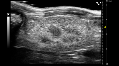
Kidney in a Small Canine
![Kidney in a small canine.jpg](/sites/default/files/styles/large/public/media-files/Kidney%20in%20a%20small%20canine.jpg.webp?itok=7Dysww1Q)
![Kidney color Doppler in small dog.jpg](/sites/default/files/styles/large/public/media-files/Kidney%20color%20Doppler%20in%20small%20dog.jpg.webp?itok=6gCHYl1m)
Kidney Color Doppler in Small Dog
![Kidney color Doppler in small dog.jpg](/sites/default/files/styles/large/public/media-files/Kidney%20color%20Doppler%20in%20small%20dog.jpg.webp?itok=6gCHYl1m)