
By Dr. Kristiina Aasa, Applications Scientist.
Here at FUJIFILM VisualSonics, we tend to throw around a lot of technical terms and acronyms about our ultrasound technology that might be confusing for those beginning to use our Vevo systems or new to ultrasound imaging in general. For this Vevo Blog entry I thought I would go back and talk about some basics of ultrasound technology. For the sake of brevity I will keep this very general.
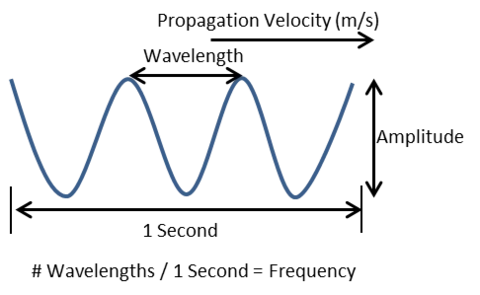
The principles of ultrasound imaging rely on the physics of sounds waves. Like all waves, there are four important properties of sound: frequency, velocity of propagation, wavelength and amplitude.
Sound waves are called ultrasound when they have a frequency above the range of human hearing, 20 kHz (or 20 000 cycles/s). Conventional ultrasound in the clinic uses frequencies from 1-20 MHz (1-20 million cycles/sec). In contrast, our Vevo Ultra High-Frequency Ultrasound refers to frequencies from 15-50 MHz.
As frequency gets higher, wavelength gets shorter and the ability to differentiate between objects (resolution) increases. Higher frequencies will attenuate (lose signal strength) more causing less depth of penetration.
Generation of an image requires transmission of the ultrasound waves from the transducer into the body (or object of interest). Propagation of the waves through the body is affected by the acoustic impedance of the particular tissues being imaged, which is determined by the density and the speed of sound in the material. A difference in acoustic impedance between adjacent tissues results in reflection of the ultrasound waves at their boundary. For example, the acoustic impedance of water is 3500 times greater than air, which means any water-air boundaries will cause reflection of the ultrasound signal. This is why air bubbles in ultrasound gel can be such a menace and cause artifacts in your image.
These reflections are detected by the transducer and are the basis of how ultrasound images are created. Differences in acoustic impedance can also cause the ultrasound waves to refract (veer off of their original trajectory) which, amongst other things, can also cause image artifacts. If the object is smaller than the wavelength of the signal (like red blood cells) it can cause the wave to scatter in various directions. As the signal travels through the body it also becomes attenuated (the signal strength decreases) due to reflection, refraction, scattering and absorption by the tissue.
There is certainly a great deal more that goes into how ultrasound waves are generated, detected and transformed into the image we see, but I will save all that for another day!