Submitted by Sam Lim, Postdoctoral Fellow, Princess Margaret Cancer Centre.
Accurate endoscopic detection of dysplasia in patients with Barrett’s esophagus (BE) remains a major clinical challenge. The current standard is to take multiple biopsies under endoscopic image guidance, but leaves majority of the tissue unsampled, leading to significant risk of missing dysplasia. Furthermore, determining if there is submucosal invasion is essential for proper staging. Hence, there is a clinical need for a rapid in vivo wide-field imaging method to identify dysplasia in BE, with the capability to image beyond the mucosal layer. We conducted an ex vivo feasibility study using photoacoustic imaging (PAI) in patients undergoing endoscopic mucosal resection (EMR) for known dysplasia. The objective was to characterize the esophageal microvascular pattern, with the long-term goal of performing in vivo endoscopic PAI for dysplasia detection and therapeutic guidance.
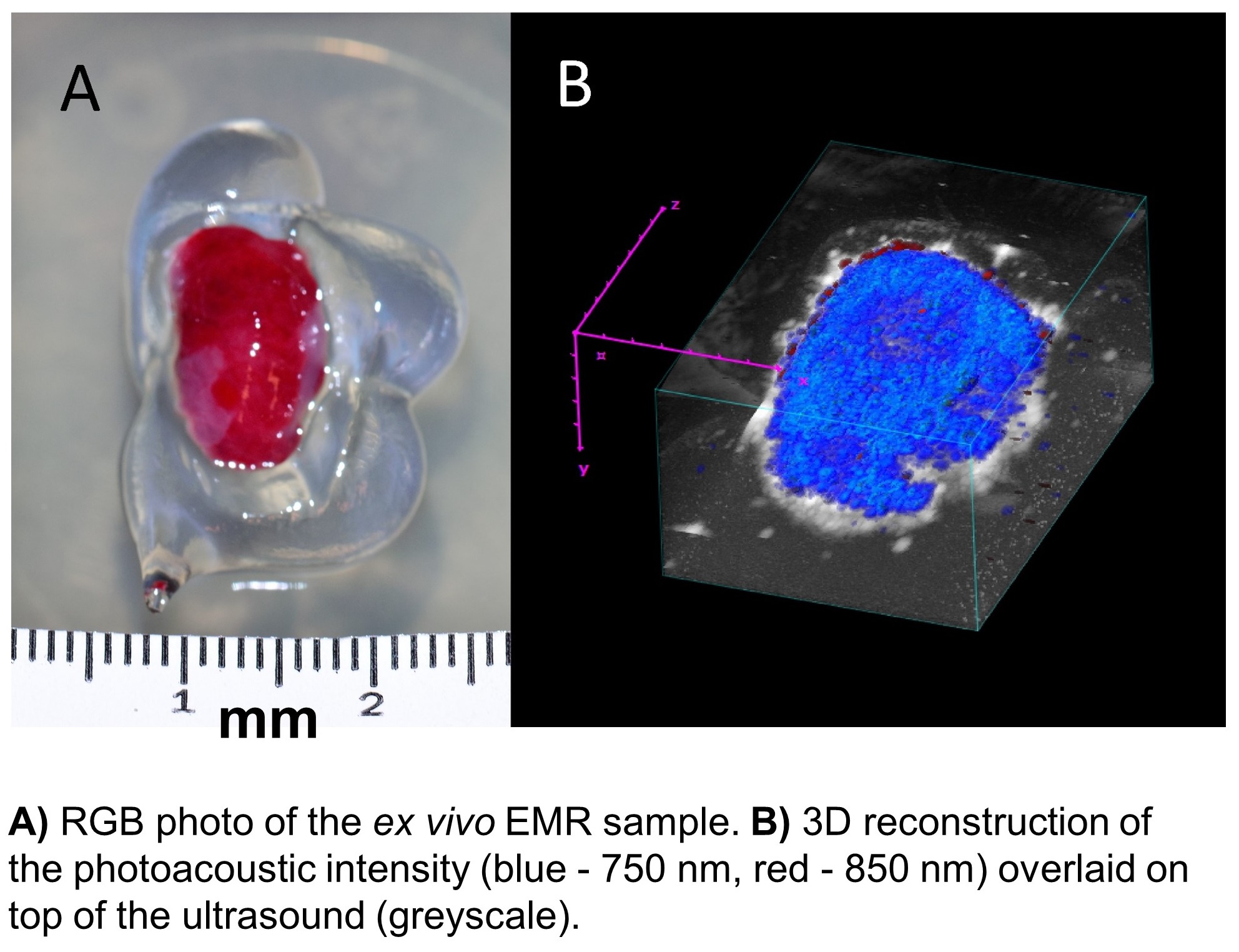
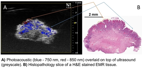
We used the VevoLAZR system to acquire 3D tomographic photoacoustic images on ex vivo human EMR tissue samples. Tissues were then sliced and fixed in formalin for histopathology with H&E staining. Our analysis consisted of co-registration of the photoacoustic images with corresponding pathologist-classified histological images. We conducted mean difference test of the total hemoglobin distribution between tissue classes. Based on our ex vivo data, changes in total hemoglobin content from intrinsic PAI (i.e. without exogenous contrast) can differentiate BE from squamous esophageal mucosa. We are investigating the diagnostic potential of PAI+antibody-targetting nanopartcles in diagnosing dysplasia from non-dysplastic BE.