
Submitted by Jing Wu, Postdoctoral Research Fellow, The University of Iowa.
Using the Cardiac Module on the Vevo 2100 system, we first determined cardiac output, left ventricular mass, and other cardiac parameters associated with high blood pressure in a mouse model of salt sensitive hypertension. These measurements were very informative and promoted us to further explore the renal perfusion in our transgenic mice. Renal perfusion is governed by blood flow in the renal artery and is a key factor determining kidney function.
Using the Vascular Module on the Vevo 2100 system, we measured the arterial luminal diameter and blood flow velocity in the right renal artery, and calculated renal blood flow using these parameters as indicated in the Figure. To our knowledge, we are one of the few centers in the United States that have successfully measured renal blood flow in transgenic mouse models using the Vevo 2100 system. We will present these exciting results at the 2018 Experimental Biology meeting. Our results demonstrate the power and versatility of the Vevo system.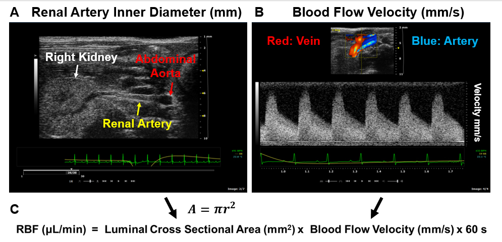 Figure: Measurement of Renal Blood Flow. A) A representative image showing the anatomical relationship of the aorta, the right kidney and the renal artery. The inner diameter of the renal artery was measured. B) Representative images of the blood flow velocity in the right renal artery during each cardiac cycle. Because the Doppler probe was placed pointing to the kidney, the renal vein was shown in red (blood flow towards the probe) and the artery was shown in blue (blood flow away from the probe). Arterial waves were originally below baseline and were reversed to above baseline for viewing and analysis. Green lines: electrocardiogram; Yellow lines: respiration. C) The renal blood flow (RBF) was calculated based on mean blood flow velocity in the cardiac cycle and the luminal cross sectional area of the renal artery in a full minute. A, area; r, radius.