
In this study, Ramasawmy et al.1 sought to compare 9 Tesla (T) MRI, Benchtop (1T) MRI, Vevo US and BLI in the ability to monitor and detect tumor growth in a xenograft colorectal metastasis model in the mouse liver.
The ability to assess successful tumor engraftment and measure tumor growth longitudinally is an extremely valuable tool in preclinical oncology research, especially when attempting to translate these results to the clinic.
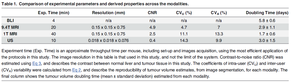
Article Summary:
- 9T MRI was considered the gold standard imaging modality for this paper
- Imaging performed on 4-5 time-points from 2-28 days post tumor engraftment
- Tumor burden measured with US and 1T MRI were highly correlated with 9T MRI; BLI overestimated tumor burden
- Tumor detection is more sensitive with US than 1T MRI at early time-points
- BLI was highest throughput; 1T MRI was lowest
- 9T MRI has lowest inter and intra-user variability (variability similar for 1T MRI vs US)
- Coefficient of variability measuring tumors manually with calipers is even worse (measured elsewhere, references provided)
- US has best resolution and is most sensitive in detecting tumor doubling time
- BLI not sensitive to variability in tumor size
Conclusion:
This study concludes that 1T MRI and US are both good options for characterizing tumor growth. A multi-modal approach might be best, using BLI to screen for successful tumor engraftment and US as a “…low-cost, and rapid platform for tumor burden assessment…”.
1. Ramasawmy R, Johnson SP, Roberts TA, et al. Monitoring the Growth of an Orthotopic Tumour Xenograft Model: Multi-Modal Imaging Assessment with Benchtop MRI (1T), High-Field MRI (9.4T), Ultrasound and Bioluminescence. PLoS One. 2016;11(5):e0156162. doi:10.1371/journal.pone.0156162.