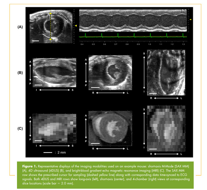
This recent article by Damen, et al. showcases the use of 4-dimensional ultrasound (4DUS) imaging for cardiac function evaluations, and compared the results to two standard techniques: short-axis M-mode (SAX MM) and cine magnetic resonance imaging (MRI).
Article Summary:
- High-frequency ultrasound (SAX MM) and MRI are currently two of the most widely used imaging techniques for non-invasive longitudinal assessment of cardiac functions in murine models.
- There are limitations to each method: SAX MM calculations require geometric assumptions to be made leading to inaccuracies, while MRI is more costly and required longer image acquisition time.
- Here an automated 4D imaging technique is implemented on the Vevo 3100 system, by acquiring high-frame rate (300 fps) cardiac- and respiratory-gated cines, then spatiotemporally compiling them into a 4D data set.
- 4DUS data was acquired in both wild type and mice with left ventricular hypertrophy, and compared with SAX MM and cine MRI.
- Quantified metrics of cardiac function included EDV, PSV, EF, SV and LVM.
- 4DUS and MRI showed high agreement in all of the assessed cardiac parameters, while SAX MM on average overestimated these values.
Conclusion:

Image credit: TOMOGRAPHY, December 2017, Volume 3, Issue 4:180-187
DOI: 10.18383/j.tom.2017.00016. Shared under Creative Commons License.
Reference:
Damen, F. W. et al. High-Frequency 4-Dimensional Ultrasound (4DUS): A Reliable Method for Assessing Murine Cardiac Function. Tomography 3, 180–187 (2017).
Available from: http://www.tomography.org/volume-3/issue-4-december/advances-in-brief/j-tom-2017-00016