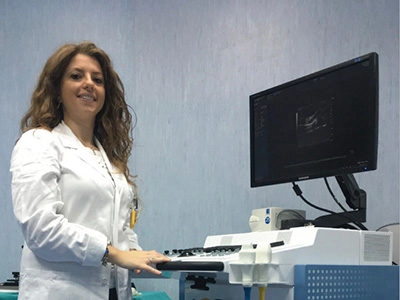
A conversation with Dr. Francesca IACOBELLIS: a specialist radiologist at Centre of Biotechnology of “A. Cardarelli” Hospital, Naples, explained how Ultra high frequency ultrasound imaging is enabling non-invasive morphological and functional assessment of biological structures in a preclinical environment.
Lots of imaging technology are widely used in preclinical research to provide, for example, cardiotoxicity, biomarker or drug expression as well as tumor behaviour. Ultra high frequency ultrasound is reliable, easy to access and perfectly adjusted to in-vivo longitudinal studies. Francesca explained the benefits of this technique.
I work as a radiologist at the Centre of Biotechnology of “A. Cardarelli” Hospital, performing preclinical research in Professor Roberto Grassi’s group of the University of Campania “Luigi Vanvitelli”, as part of my PhD studies. Ultra high frequency (UHF) ultrasound allows me to simultaneously obtain in vivo anatomical, functional, and physiological data in real time, with a resolution down to 30 µm. A big advantage of this technology is that I can perform non-invasive longitudinal studies of animal models, which is a more ethically acceptable way of undertaking preclinical research.
I have been using the VisualSonics technology since 2009, starting with cardiovascular imaging and moving on to abdominal applications. The Vevo system is particularly suitable for cardiovascular applications, offering exceptional quality images with high temporal resolution, which is ideal for the high cardiac frequency of small animals.
B-mode ultrasound allows the heart morphology and function to be studied, complemented by M-mode assessment, and colour and power Doppler analysis of cardiac blood flow.
The Vevo system has played an important role in several projects.