
A conversation with Dr Florian RAES, a former PhD student at the Centre d’Imagerie du Petit Animal (CIPA), PHENOMIN-TAAM in CNRS Orléans, now Post-Doctoral Research Fellow, Preclinical Molecular Imaging team, The Institute of Cancer Research, London.
Bioluminescence and near-infrared fluorescence imaging are widely used in preclinical oncology research to provide information about tumor proliferation and biomarker expression, for example. However, these techniques have their limitations, and additional examinations using complementary technologies are often required. Dr Florian Raes, shares the development of an all-in-one multimodal imaging platform combining the Vevo® LAZR’s ultrasound and photoacoustic capabilities with bioluminescence and near-infrared fluorescence.
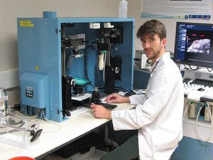
When Dr Florian Raes joined CIPA, a facility dedicated in imaging services and R&D for academia and industry, the laboratory had just purchased a Vevo LAZR. This high frequency ultrasound and photoacoustic imaging system enables the detection of tumors and determination of the precise location of metastases in the body, supplementing data from bioluminescence, nearinfrared fluorescence and conventional ultrasound investigations. Dr Raes explains: “The ultra high frequency ultrasound offers superior resolution down to 30 μm, with a maximum depth of 4 cm, giving 2D and 3D anatomical data, plus information on blood flow, tumor perfusion status and vascular biomarkers. This is complemented by a very sensitive photoacoustic imaging modality, enabling the assessment of tumor oxygenation status and, with the aid of contrast agents, molecular imaging.
“We realized that there was considerable benefit in integrating all four imaging technologies – photoacoustics, high frequency ultrasound, bioluminescence and near-infrared fluorescence – into one system. This new hybrid platform makes
it possible to do everything in one go. It’s a big advantage for translational research.” ~ Dr. Florian Raes
Initially, the focus was on photoacoustics. We used it successfully for tumor hypoxia studies in animal models with pancreatic and mammary malignancies, and for the investigation of the effects of some chemotherapies. Later, we began to work with the ultrasound features of the instrument and discovered the Vevo LAZR’s potential to deliver both anatomical and functional information. The system complementing the bioluminescence and fluorescence imaging systems, is now used extensively in the department.
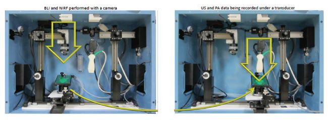
Dr. Raes states: “After some mechanical adjustments and software changes, we were able to modify the Vevo LAZR to incorporate both a transducer and a CMOS Hamamatsu scientific camera within the system’s light enclosure.” This multimodal platform combines anatomical, functional and molecular information from the Vevo LAZR with high sensitivity tumor proliferation and biomarker expression measurements using bioluminescence and near-infrared fluorescence imaging, improving characterization of key tumor biomarkers.
Multimodal imaging systems can also minimize the need to repeatedly administer anaesthetic to animals and transfer them between different imaging devices, making it less traumatic for the animal and allowing physiological parameters – such as the heart rate, respiration rate and body temperature – to be monitored in real time. Moreover, this platform allows rapid examination of the animal, which remains in the same bed, making data analysis and comparison of matching images much easier.
Dr. Raes reports: “There are other benefits too; bioluminescence is commonly used to detect metastases, but light diffused from the primary tumor can mask small growths in the surrounding tissues. Complementing this technique with ultrasound and photoacoustic imaging. We realized that there was considerable benefit in integrating all four imaging technologies – photoacoustics, high frequency ultrasound, bioluminescence and near-infrared fluorescence – into one system. This new hybrid platform makes it possible to do everything in one go. It’s a big advantage for translational research.”
Bioluminescence and near-infrared fluorescence imaging are widely used in preclinical oncology research to provide, for example, information about tumor proliferation and biomarker expression. However, these techniques have their limitations, and additional examinations using complementary technologies are often required. Dr Florian Raes, shares the development of an all-in-one multimodal imaging platform combining the Vevo® LAZR’s ultrasound and photoacoustic capabilities with bioluminescence and near-infrared fluorescence can enable visualization of metastases that might otherwise not be seen due to the poor resolution of in vivo bioluminescence methods.
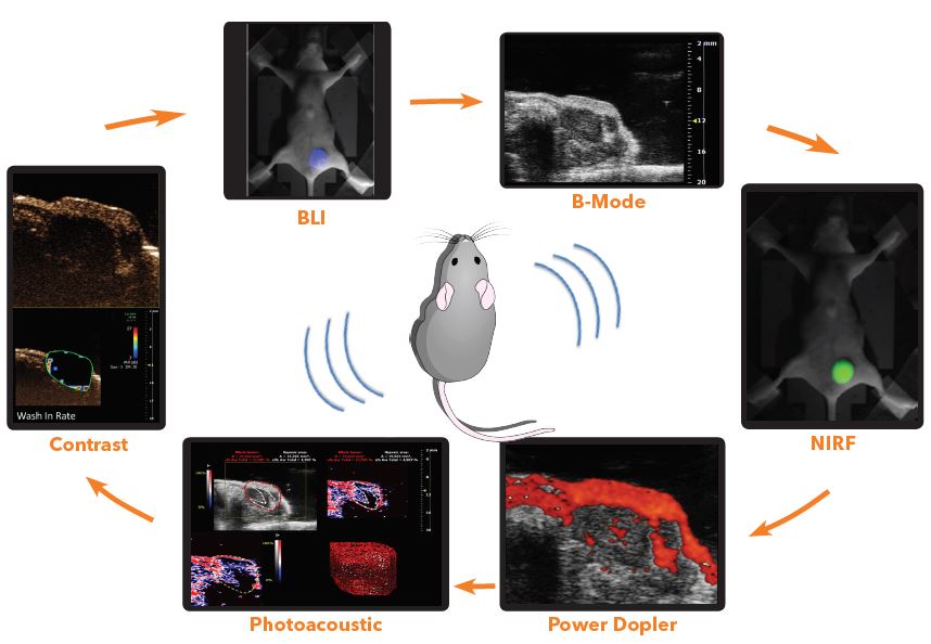
 Once detected, metastases can be monitored and precisely measured by ultrasound, using 2D or 3D tools, while power Doppler allows small vessels with a slow flow to be located, and the percentage of vascularization to be determined. Photoacoustics adds another dimension, as it enables the determination of haemoglobin oxygen saturation, an indication of tumor hypoxia, in the same way that specific contrast agents can be used as biomarkers. It can also be combined with contrast-enhanced ultrasound to investigate microvascularization mechanisms in the tumor, with bioluminescence for tumor proliferation studies, and with near-infrared fluorescence for biomarker imaging. It’s a very versatile platform with great potential for a wide range of preclinical and research studies.
 By integrating all four modalities into one platform, CIPA has taken a big step forward in respect of its imaging capabilities. While researchers could have continued to perform each type of examination individually using a specific imaging technology, This new hybrid platform makes it possible to do everything in one go. It’s a big advantage for translational research.
About CIPA: The Centre d’Imagerie du Petit Animal (CIPA) is a division of UPS44 TAAM (Typing and Archiving of Animal Models), located on the National Center for Scientific Research (CNRS) campus at Orléans, member of PHENOMIN national infrastructure. Founded in 2002, its expertise deals on oncology, inflammation, drug vectorisation and phenotyping of murine models of human pathologies to support research being performed across the academic community and industry.
By integrating all four modalities into one platform, CIPA has taken a big step forward in respect of its imaging capabilities. While researchers could have continued to perform each type of examination individually using a specific imaging technology, This new hybrid platform makes it possible to do everything in one go. It’s a big advantage for translational research.
About CIPA: The Centre d’Imagerie du Petit Animal (CIPA) is a division of UPS44 TAAM (Typing and Archiving of Animal Models), located on the National Center for Scientific Research (CNRS) campus at Orléans, member of PHENOMIN national infrastructure. Founded in 2002, its expertise deals on oncology, inflammation, drug vectorisation and phenotyping of murine models of human pathologies to support research being performed across the academic community and industry.