Single Wavelength
Detect and Fuse Photoacoustic Signals with Detailed Anatomical Images
For visualization of chromophores such as hemoglobin, melanin, dyes and nanoparticles
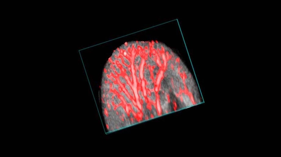
PA-Mode Single Wavelength allows the detection and fusion of photoacoustic signals with detailed anatomical images. Using a tuneable near infrared laser, the subject is illuminated at the desired wavelength (680-970nm and 1200-2000nm*) in order to visualize chromophores such as hemoglobin, melanin, dyes and nanoparticles. These signals are localized to specific anatomy as visualized through simultaneously acquired high-resolution ultrasound images.
(Shown left) Mouse ear visualized using single wavelength