Shear Wave Elastography
Shear Wave Elastography for Detecting Tissue Stiffness
All new and fully integrated on the Vevo F2!
.")
Shear wave elastography induces shear waves via ultrasound push pulse(s).
What is Elastography?
Elastography is a non-invasive imaging technique used for detecting tissue stiffness. It is an established method used for detecting and staging liver fibrosis.
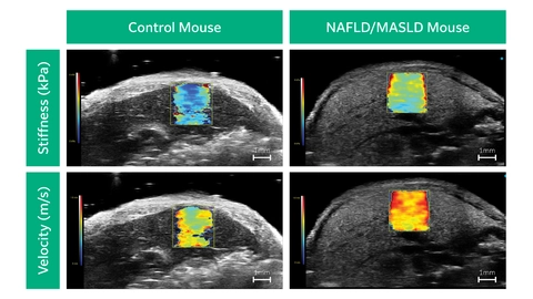
Low-frequency vibrations, or shear waves, are introduced into the tissue and detected by the UHF29x transducer on the Vevo F2 system. Tissue stiffness and shear wave velocity can then be visualized using a color map on the system, and quantified in particular regions of interest. Shear wave speed is used as a measure of tissue stiffness; faster shear wave propagation equates to a stiffer tissue.
Shear Wave Elastography on the Vevo F2
When studying disease models it is imperative to have the right tools for the model. Vevo high-resolution ultrasound is a proven and valuable tool to study small animal models of disease. The increased resolution and details are required to visualize and quantify subtle changes in these smaller models of disease. The integration of elastography with high-frequency ultrasound enables researchers to precisely visualize the anatomy of interest and quantify tissue stiffness in different sub-sections of an organ, all non-invasively.
How Preclinical Elastography Can Advance Your Research
- Visualize tissue stiffness non-invasively
- Easily quantify stiffness and shear wave velocity
- Assess liver fibrosis with high anatomical resolution
Request a Quote or Demo
Elastography in Action
Shear Wave Elastography Mode
Live Webinar
Learn From The Experts
Learn about Shear Wave Elastography and High-frequency Ultrasound from experts in the field! Dr. Jean-Luc Gennisson, Paris-Saclay University, and Dr. Gilles Renault, INSERM Cochin Institute present their research in this webinar.