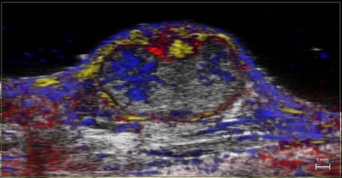
High-resolution ultrasound (greyscale) and spectrally unmixed photoacoustic (color) image of a subcutaneous tumor showing nanoparticle distribution (yellow) as well as oxygenated (red) and deoxygenated (blue) hemoglobin signal.
High-resolution ultrasound (greyscale) and spectrally unmixed photoacoustic (color) image of a subcutaneous tumor showing nanoparticle distribution (yellow) as well as oxygenated (red) and deoxygenated (blue) hemoglobin signal.