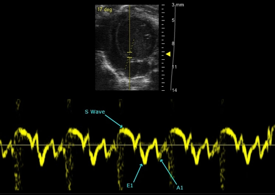
Tissue Doppler Imaging
Measure Tissue Movement Typically Performed on the Myocardium

Assess Diastolic Function and Myocardial Strain

Detect changes in tissue motion

Tissue Doppler Mode at the mitral annulus and software analysis tools allow for various measurements to be preformed, here the early (E’), atrial (A’), and systolic (S) peak velocities are measured.

Tissue Doppler Imaging allows for measurements of tissue movement, typically performed on the myocardium.

This tool is ideal to assess diastolic function of the left ventricle by placing a sample volume at the mitral annulus, where changes in tissue motion are often detected prior to changes in blood flow spectrums through the same valve.

Ideal For:

  • Diastolic Dysfunction
  • Heart Failure
  • Load-Independent Tissue Measurements