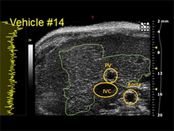
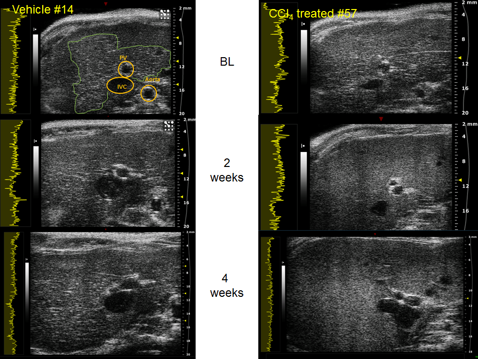
Ultrasound image of normal and treated rats is shown in its transverse view. CCl4 is used to induce fibrosis (details in the webinar). Ultrasound in this longitudinal study was conducted at the intervals shown in the image below. The images in the column for the vehicle shows annotations in the first image to guide your understanding of this ultrasound image. The green trace shows the area of the liver that was investigated. Note the normal liver tissue within this trace can be described as dense grainy and hyperechoic signal. This characteristic is lost in the CCl4 treated mice at 2 weeks. Terri Swanson describes how she acquires this image with reproducibility and consistency.
Submitted by Terri A. Swanson, Senior Scientist, Comparative Medicine, Pfizer Inc. and Theresa Tuthill, Head of Cardiovascular, Metabolic, and Musculoskeletal Imaging for the Clinical and Translational Imaging group at Pfizer Inc