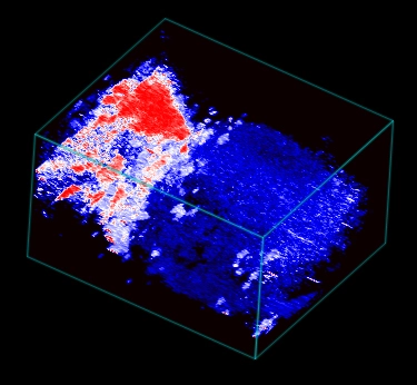
3D rendered photoacoustic image of the mouse hindlimb showing a parametric map of oxygen saturation with red denoting higher sO2 values. Application of a tourniquet made of tubing was used to restrict blood flow into the distal part of the limb. Much lower sO2 is seen in the distal part of the limb during this acute ischemia.